Nature Knows and Psionic Success
God provides
The Science Behind Echoic Memory: How Sound Lingers in Your Mind

This Is Why We Remember Certain Sounds
Medically reviewed by Shaheen Lakhan, MD, PhD, FAAN
Echoic memory, also known as auditory sensory memory, involves the short-term recall of sounds you’ve just heard. The memory of a sound might linger in your mind very briefly after the actual auditory stimulus has ended. It is a bit like an echo of a sound that exists only in your mind.
The brain utilizes several different types of memory , and echoic memory serves an essential purpose. While it is very short, lasting around four seconds, it allows us to temporarily store the sound until it can be processed. tl;dr
We’ve all heard a sound before that seems to stick out in our minds. Maybe it’s a ringing noise or a honking noise. Perhaps it’s the sound of your cat’s meow or dog’s bark. Whatever the noise, it seems to linger in your brain long after the sound has stopped. This is what’s known as echoic memory.
Unlike images that you can repeatedly go back and look at, you cannot do that with sound (unless it’s recorded of course). This is where echoic memory comes in because it allows us to recall sounds even when we’re no longer in earshot of them.
There are a few factors that can influence your auditory sensory memory and it may be possible to even improve our echoic memory. What Exactly Is Echoic Memory?
Echoic memory is defined as a type of sensory memory that temporarily stores auditory information. This serves an essential purpose: it allows a sound to be stored just long enough to be processed and understood.
During the 1970s, researchers discovered that auditory information will disappear from memory after about five seconds—unless you pay attention to it. By focusing your attentional spotlight on the sounds, the information will more likely make its way into short-term memory .
What makes echoic memory so important? Unlike visual information, which the viewer can look at often for as long as they want and be reviewed when needed, sounds are fleeting. They are presented once and usually cannot be re-experienced unless an audio recording exists. Takeaway
By having echoic memory, people are able to briefly hold on to that sound so that it can then be processed and transformed into meaning. How Does Echoic Memory Really Work?
According to one model, sensory memory is the first stage of memory . At any given moment, you are taking in sensory information about the world around you. Because there is no way to focus on all of the different details of every sensation you experience, your brain creates a snapshot of your sensory experience. This allows you to then focus on details that you might have missed.
In the case of echoic memory, this allows you to retain a brief impression of an auditory sensory experience even after the original stimulus has ended or disappeared. Then, by attending to these details, you can transfer important information into the next stage of memory, known as short-term memory. Takeaway
Echoic memory is automatic, meaning it happens without having to make a conscious effort.
After a noise is produced, the sound waves are picked up by the human ear, where they affect the auditory nerve. This turns the sound waves into electrical impulses transmitted to the brain.
Once the sound reaches the brain, an echoic memory is formed. The brain processes this information and then stores it in the primary auditory cortex (PAC) on the opposite side of the brain that receives the sound.
So if your right ear received the sound, the echoic memory for that sound would be stored in the primary auditory cortex in the left hemisphere of the brain. Sounds are often received by both ears, meaning the echoic memory is stored in both hemispheres.
The brief storage in echoic memory gives the brain time to interpret the sound and determine its characteristics. The sound may be transferred into working memory for further interpretation.
Information also cannot be retained in echoic memory through rehearsal. Subsequent sounds are also continually displacing the previously heard information. This ever-updating nature enables echoic memory and other types of sensory memory to act as real-time monitors for new information in the environment. Duration and Capacity of Echoic Memory
Echoic memory is an important part of your experience of the world, allowing you to store auditory information long enough that you can process and understand it. Takeaway
Echoic memories are very brief, lasting in the auditory storage system for approximately two to four seconds.
Brain imaging technology has also allowed researchers to learn more about how auditory sensory memory works. In one study, researchers found that after a sound stimulus, activity occurs in a portion of the auditory cortex and lasts around two to five seconds after the sound. Echoic Memory vs. Iconic Memory
However, echoic memory lasts longer than iconic memory , which is the ultra-short memory of visual imagery. Where a sound might linger in your echoic memory for up to four seconds, your ability to store visual information lasts for just a few hundred milliseconds.
While iconic memory is incredibly short, visual imagery is more enduring. In most cases, you can spend time looking at visual stimuli for longer periods, or you may even be able to view it repeatedly.
A sound, on the other hand, is often only produced once. Depending on the source of the sound, you may never be able to experience it again. This is why echoic memory is so important.
Echoic memory allows you to briefly hold on to this aural information to fully understand it, even after the original source is gone. Examples of Echoic Memory Some examples of how echoic memory is used include: Listening to music : As you listen to music, your brain briefly recalls the previous sounds, creating a connected and continuous experience that allows you to recognize the many notes as a cohesive song. Environmental noises : Echoic memory can also help you make sense of the noises you hear each day in the world around […]
The best adaptogenic herbs for managing stress

Advertisement Ann Vandersteel joins Mike Adams to expose government TYRANNY and TREASON against the people Steve Quayle and Mike Adams: The American government turns its weapons of WAR against the PEOPLE Advertisement
Adaptogens are herbs or active plant compounds that bolster the body’s ability to respond to stress . They can also help restore balance after stressful situations.
Stress, simply put, is your body’s normal reaction to pressure or a visible/palpable threat. According to researchers at the University of California, Berkeley , stress can sometimes be good for you as it can “push you just to the level of optimal alertness, behavioral and cognitive performance.” In fact, their study on rats showed that brief stressful events stimulated stem cells in the animals’ brains to become new nerve cells, which improved the rats’ mental performance.
When faced with a stressor, the human body is said to react very swiftly and very efficiently . Because physiological changes mediated by the sympathetic nervous system, which releases stress hormones, happen so quickly, people in stressful situations often find themselves reacting before they can even think about what they’re doing. Once the trigger is gone, the parasympathetic nervous system presses the brakes on your stress response.
But if you are constantly subjected to everyday pressures or other stressful triggers, your parasympathetic nervous system may be hindered from dampening your stress response. Chronic low-level stress causes physiological changes that can inadvertently lead to various health issues. Problems linked to chronic stress include fat buildup and weight gain and increased blood pressure, which can ultimately damage your blood vessels and raise your risk of heart attack or stroke.
Fortunately, there are plenty of ways to manage your stress naturally . Aside from practicing relaxation and mindfulness techniques, you can take adaptogenic herbs in the form of supplements or tea to help your body cope with stress in the most optimal way possible. On top of being safe, adaptogenic herbs can help reduce your stress response when it’s no longer needed so your body can achieve a state of internal balance and stability known as homeostasis. 5 Adaptogenic herbs for improving stress resilience
Plenty of herbs used in ancient medicinal systems are still used today to treat various health issues. Although only a few medicinal herbs can be called adaptogens, these herbs are relatively common and easy to find, so they’re readily available to anyone in need of non-pharmaceutical stress support. Medicinal herbs are known for having a variety of health-supporting properties, so expect more than just one health benefit when you use adaptogenic herbs.
If you need help managing stress or optimizing your stress response, try these five adaptogenic herbs : Asian ginseng ( Panax ginseng )
Chronic stress is linked to an increased risk of heart disease because of the effects of cortisol. Having high levels of this stress hormone raises not only blood cholesterol and triglyceride levels , but also blood sugar and blood pressure.
High cortisol levels also have a negative impact on your immune system. While a temporary increase in cortisol can boost your immune response, persistently high cortisol levels can lead to chronic inflammation , which can be damaging to tissues and organs. Long-term stress has also been found to weaken your immune system as it decreases the number of circulating white blood cells , the immune cells that help fight infections.
According to a study published in the Journal of Ginseng Research , Asian ginseng exerts its adaptogenic effects by helping stimulate or suppress immune response as well as regulating blood vessel function and blood pressure. Thanks to its immunomodulatory, vasomodulatory and cardioprotective properties, Asian ginseng can promote vitality, enhance physical performance and increase your body’s resilience to stress. American ginseng ( Panax quinquefolius )
American ginseng is a species of ginseng native to North America. Like Asian ginseng, this herb is considered a powerful adaptogen that offers benefits for the brain and immune system. (Related: Four of the best adaptogens that help you beat stress .)
According to studies, American ginseng contains plenty of antioxidant compounds called ginsenosides. These bioactive compounds not only protect the brain from oxidative stress but also support the production and growth of new neurons . The ginsenosides in American ginseng have also been found to support optimal mental clarity, improve attention and memory , and reduce mental fatigue.
The memory-boosting effects of American ginseng can be particularly helpful to students under academic stress. Although stress is thought to enhance memory formation, studies show that it actually impairs memory retrieval and hampers your brain’s ability to update memories with new information. Supplementing with American ginseng can therefore help improve memory performance in times of stress.
Like Asian ginseng, American ginseng also exerts immunomodulatory effects, thanks to bioactive polysaccharides. Depending on your body’s needs, these polysaccharides can either stimulate your immune system or suppress immune response to protect your body from chronic inflammation. According to research, American ginseng contains different classes of polysaccharides, which constitute about 10 percent of the dried root’s weight. Arctic root ( Rhodiola rosea )
Arctic root, also known as golden root or roseroot, has a long history of use in traditional medicine. A popular remedy in many parts of Europe and Asia, arctic root is known for its ability to stimulate the nervous system , enhance physical performance and treat stress-induced fatigue and depression. (Related: Rhodiola significantly improves mental health and promotes a calm state of mind .)
Pre-clinical and clinical trials show that, as an adaptogen, arctic root has brain-stimulating and calming properties that can help improve memory performance and mood during times of stress. A Russian-Swedish study involving 40 medical students reported that those who supplemented with arctic root extract during examination week enjoyed a boost in physical and mental performance , significant reductions in mental fatigue, greater mood stability and an increase in motivation to study.
A separate study by Swedish researchers also reported on arctic root’s effectiveness in alleviating fatigue. Supplementing with arctic root daily for 28 days was found to benefit burnout patients by significantly reducing fatigue […]
Brain implants can help people with paralysis ‘speak’ through screens faster, more accurately than before, new studies show
Noah Berger/UCSF A participant in a study of speech neuroprostheses uses a digital link wired to her cortex to interface with an avatar. By Jacqueline Howard, CNN
(CNN) — Dr. Jaimie Henderson had a single wish throughout childhood: for his father to be able to speak with him. Now a scientist and neurosurgeon at Stanford Medicine, Henderson and his colleagues are developing brain implants that might be able to make similar wishes come true for other people with paralysis or speech impairments.
Two studies published Wednesday in the journal Nature show how the brain implants, described as neuroprostheses, can record a person’s neural activity when they attempt to speak naturally, and that brain activity can then be decoded into words on a computer screen, through audio speech or even communicated using an animated avatar.
“When I was 5 years old, my dad was involved in a devastating car accident that left him barely able to move or speak. I remember laughing at the jokes he tried to tell, but his speech ability was so impaired that we couldn’t understand the punchline,” Henderson, an author of one of the studies and professor at Stanford University, said in a news briefing about his research.
“So I grew up wishing that I could know him and communicate with him,” he said. “And I think that early experience sparked my personal interest in understanding how the brain produces movement and speech.”
Henderson and his colleagues at Stanford and other US institutions examined the use of implanted brain sensors in 68-year-old Pat Bennett. She had been diagnosed with amyotrophic lateral sclerosis in 2012, and it affected her speech.
The researchers wrote in their study that Bennett can make some limited facial movements and vocalize sounds but is unable to produce clear speech due to ALS, a rare neurological disease that affects nerve cells in the brain and spinal cord.
In March 2022, Henderson conducted the surgery to implant arrays of electrodes in two areas of Bennett’s brain. The implants recorded neural activity as Bennett attempted to make facial movements, make sounds or speak single words.
The arrays were attached to wires that came out of the skull and were connected to a computer. Software decoded the neural activity, converting the activity into words that were displayed on the computer screen in real time. When Bennett finished speaking, she pressed a button to finalize the decoding.
The researchers evaluated this brain-to-computer interface with Bennett attempting to speak with vocalizations and to only “mouth” words with no vocalization.
With a 50-word vocabulary, the rate of errors in the decoding was 9.1% on the days Bennett vocalized and 11.2% on the silent days, the researchers found. When using a 125,000-word vocabulary, the word error rate was 23.8% across all vocalizing days and 24.7% for silent days.
“In our work, we show that we can decipher attempted speech with a word error rate of 23% when using a large set of 125,000 possible words. This means that about three in every four words are deciphered correctly,” Frank Willett, an author of the study and Howard Hughes Medical Institute staff scientist affiliated with the Neural Prosthetics Translational Lab, said in the news briefing.
“With these new studies, it is now possible to imagine a future where we can restore fluent conversation to someone with paralysis, enabling them to freely say whatever they want to say with an accuracy high enough to be understood reliably,” he said.
The researchers say the decoding happened at high speeds. Bennett spoke at an average pace of 62 words per minute, which “more than triples” the speed of previous brain-computer interfaces that had about 18 words per minute for handwriting models.
“These initial results have proven the concept, and eventually technology will catch up to make it easily accessible to people who cannot speak,” Bennett wrote in a news release . “For those who are nonverbal, this means they can stay connected to the bigger world, perhaps continue to work, maintain friends and family relationships.”
Yet for now, the researchers wrote that their findings are a “proof of concept” that decoding speaking movements with a large vocabulary is possible, but it needs to be tested on more people before it can be considered for clinical use.
“These are very early studies,” Willett said. “And we don’t have a big database of data from other people.” ‘There is hope’
The other study published Wednesday involved a woman who could not speak clearly due to paralysis after she had a stroke in 2005, when she was 30 years old. In September 2022, an electrode device was implanted in her brain at UCSF Medical Center in San Francisco, with no surgical complications.
The implant recorded neural activity, which was decoded into text on a screen. The researchers wrote in the study that they found “accurate and rapid large-vocabulary decoding” with a median rate of 78 words per minute and a median word error rate of 25%.
Separately, as the patient attempted to silently speak, her neural activity was synthesized into speech sounds. The researchers also developed an animation of a facial avatar to accompany synthesized speech, based on the patient’s attempted facial movements.
“Faster, more accurate, and more natural communication are among the most desired needs of people who have lost the ability to speak after severe paralysis,” the researchers, from the University of California, San Francisco and other institutions in the United States and United Kingdom, wrote in their study. “Here we have demonstrated that all of these needs can be addressed with a speech-neuroprosthetic system that decodes articulatory cortical activity into multiple output modalities in real time.”
The two new brain implant studies are “overlapping” in their findings and their “long-term goal” to restore communication for people with paralysis, Dr. Edward Chang, a neurosurgeon and author of the study from UC San Francisco, said in the news briefing.
“The results from both studies — between 60 to 70 words per minute in both of them — is a real milestone for our field, in general, and we’re really excited about it because it’s coming from […]
Researchers to probe brain mechanisms behind free will
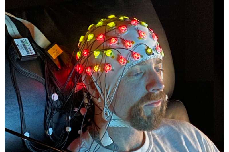
EEG equipment being used to monitor brain activity. Credit: George R. Mangun Picture this scenario: You and a friend are walking around your neighborhood when you stop at a crosswalk. As you wait, the noises of the world and your internal thoughts all vie for your attention. Suddenly, you see a motorist nearly hit a bicyclist.
“Whoa, did you see that?” you say to your friend.
“I sure did; that was a fully restored 1967 Ford Mustang,” your friend replies, referring to a car separate from the near-traffic collision.
Despite being in the same place at the same time, and looking at the same scene, you and your friend paid attention to different things. Why? And how?
One possibility is that different things in the environment attracted your attention differently. But another possibility is that you and your friend made voluntary decisions about what to pay attention to, exerting your free will accordingly.
Distinguished Professor George R. Mangun, director of the UC Davis Center for Mind and Brain, is launching a project to better understand the cognitive mechanisms behind realistic voluntary attention, or attention directed by an individual’s free will. The project will be conducted in collaboration with engineering colleagues at the University of Florida.
“How we use free will to focus attention influences our momentary awareness and our consciousness,” said Mangun.
The role of voluntary attention isn’t just important to understanding healthy brain function, he said. It’s also critical to understanding disorders of the brain.
“Almost every neurological or psychiatric disorder touches some component of attention, and this sometimes involves deficits in free will, such as in Parkinson’s disease,” he said. Free will without the ‘free’
For decades, neuroscientists have probed the cognitive and neural mechanisms behind voluntary attention by using external cues (simple stimuli such as arrows) to direct their research subjects’ attention to specific locations in front of them. But this is a far cry from how human voluntary attention naturally occurs.
In the real world , our free will enables us to control what we pay attention to, even in the face of potential distractions. To accommodate for this reality, neuroscientists often investigate the concept of free will through voluntary motor actions, asking study participants to push a button of their own volition without prompting from a researcher.
Mangun’s project will bring these two lines of research together. Because orienting attention is a cognitive act and not a motor one, the researchers hope to take an important step forward in understanding both free will and voluntary attention. Measuring true ‘free will’
Mangun’s project will build upon previous experiments his lab conducted analyzing the neural activity that precedes voluntary decision-making.
In a study published in the Journal of Cognitive Neuroscience in 2014, Mangun and his colleagues recount experiments in which they asked participants to look at a spot in the center of a computer screen. They then prompted the participants to focus attention somewhere else on the screen using free will to choose where but doing so without moving their eyes to look at the attended location directly.
By making this request, the researchers separated the cognitive act of attending from the motor act of moving the eyes to look. Mangun and his colleagues recorded the participants’ brain waves (electroencephalography or EEG signals), both before and after they used free will to focus attention.
“It turns out that the ongoing pattern of brain activity preceding the subject’s free will choices could predict where they were going to later focus their attention,” Mangun said. “Using neuroscience methods, we were able to do a sort of mind reading.” Improving prosthetics and diversifying research
Mangun’s new work takes the next step forward by applying machine learning approaches and advanced brain imaging methods to pinpoint the precise neural underpinnings of these predictive brain signals. The highly interdisciplinary project brings together engineers such as postdoctoral scholar Sreenivasan Meyyappan and psychology doctoral candidate John Nadra.
Probing and refining the cognitive mechanisms behind voluntary attention could help improve technological interventions, like prosthetics. Currently, many prosthetics harness peripheral information like muscle activity. This new research could shift those signals from the muscle to the brain itself.
“You can train a person to use a mechanical arm by tapping into the electrical activity of the shoulder muscles and then they learn to contract those muscles to drive that mechanical arm,” said Mangun. “But in addition, one can use the brain’s electrical activity, for example in the motor cortex , directly to do the same. Our research may open doors to tapping into brain signals even earlier in the chain of neural events.”
In addition to its potential broad impacts on health and wellness, the project will open doors to students interested in conducting neuroscience research.
In the grant application, Mangun noted that the project will help advance and diversify STEM education by involving undergraduates, graduates and postdoctoral scholars, recruiting students through the UC Davis Summer Undergraduate Research Program and the Young Scholars Program .
“One of the great benefits of studying at a research-intensive university like UC Davis is the possibility for students to participate in cutting-edge research—it’s part of who we are,” he said.
Provided by UC Davis
Study: Irregular sleep patterns linked to harmful gut bacteria

Advertisement
A new study has associated irregular sleep patterns to “bad” gut bacteria .
The study conducted by researchers from King’s College London (KCL) and ZOE, the personalized nutrition company, is the first to discover several links between social jet lag, defined as “the shift in your internal body clock when your sleeping patterns change between workdays and free days,” and diet quality, dietary habits, inflammation and gut microbiome composition in a single cohort.
Details of the study were published in the European Journal of Nutrition.
Earlier studies have revealed that working shifts disrupts the body clock and can increase the risk of weight gain, heart problems and diabetes in certain people.
However, not much is known about how human biological rhythms can be affected by smaller inconsistencies in sleeping patterns, such as when you wake up early with an alarm clock on workdays, compared to when you wake naturally on non-workdays, especially if you are working regular hours. Major sleep disruptions can affect overall health negatively
Dr. Wendy Hall, a senior author from KCL, said major disruptions in sleep can significantly affect your health. The KCL and ZOE study is the first of its kind to show how even small differences in sleep schedules throughout the week can be linked to differences in gut bacterial species.
Hall said some of these associations were linked to dietary differences but data also suggests that other unknown factors may be involved. She added that conducting intervention trials will help researchers find out if improving sleep time consistency can offer beneficial changes in the gut microbiome and related health outcomes. (Related: Sleep deprivation linked to health issues like obesity and cognitive decline .)
The composition of the microbes in your gut microbiome may negatively or positively affect your health by producing toxins or beneficial metabolites. Certain species of microbes can correspond to someone’s risk of long-term health issues like diabetes , heart disease and obesity.
The microbiome is influenced by the food you eat, meaning you can improve the diversity of microbes in your gut .
In a cohort of 934 volunteers from the ZOE PREDICT study, the largest ongoing nutritional study of its kind, scientists analyzed blood, stool and gut microbiome samples.
The scientists also studied glucose measurements in volunteers with irregular sleep patterns compared to others who had a routine sleep schedule.
Previous research into the links between social jet lag and metabolic risk factors has been conducted in populations with obesity or diabetes, but the cohort included mainly lean and healthy people. Most of the volunteers get more than seven hours of sleep per night throughout the week.
Results revealed that there was only a 90-minute difference in the timing of the midpoint of sleep, which is the halfway point between sleep time and wake-up time, associated with differences in gut microbiome composition.
Having social jet lag was associated with lower overall diet quality, higher intakes of sugary beverages and lower intakes of fruits and nuts, which may directly affect the abundance of specific microbiota in the gut.
Researchers also reported that three out of the six microbiota species more abundant in the social jet lag group have “unfavorable” associations with health.
The microbes were linked to poor diet quality, indicators of obesity and cardiometabolic health and markers in the blood that are related to higher levels of inflammation and cardiovascular risk.
Kate Bermingham, the study’s first author from KCL and a senior nutrition scientist at ZOE, said sleep is “a key pillar of health.” She added that even a short 90-minute difference in the mid-point of sleep can encourage microbiota species that have “unfavorable associations” with your well-being.
Earlier studies have shown that social jetlag is linked to adverse health issues like chronic illness, weight gain and even mental fatigue. Tips for improving sleep quality
Try the suggestions below if you need help maintaining regular sleep patterns and improving sleep quality:
Disconnect devices one hour before bed
Devices like smartphones, tablets, or laptops can keep your brain wired, making it hard to truly wind down for bed.
The blue light from your electronic devices can also suppress your natural production of melatonin. Try setting your phone aside for at least one hour or more before going to bed.
Invest in a quality mattress and bedding
If you can’t sleep because your mattress is uncomfortable and lumpy, invest in a quality mattress for your needs and preferences.
Buy a supportive mattress and pillow so your spine gets proper support. This will also help prevent various aches and pains.Get sheets and blankets that feel comfortable to the touch and help you maintain a comfortable temperature during the night. Block out light in your room Too much light exposure can affect your sleep and circadian rhythm. Get blackout curtains for your windows or wear a sleep mask to block light and prevent it from interfering with your rest. Minimize noise If you cannot eliminate nearby sources of noise, try drowning them out with a fan or white noise machine.Use earplugs or headphones to stop various sounds from preventing you from sleeping. Make sure you get at least seven hours of sleep To ensure that you get the recommended amount of sleep each night, make an effort to build that time into your schedule.Think of your desired wake-up time, then try to find the best bedtime that lets you get at least seven hours of sleep.If possible, give yourself a couple of extra hours before bed so you can prepare before your bedtime. Set your alarm for the same time every day It can be hard for your body to get used to a healthy sleep routine if you keep waking up at different times.Choose a wake-up time and follow it regularly, even on weekends or on days when you don’t have work.Visit Health.news for more articles with tips on how to improve your sleep quality, gut health and overall well-being.Watch the video below to learn how acai berries can support a healthy immune system and healthy sleep patterns . Use acai berries to support a healthy immune […]
Researchers identify mathematical rule behind the distribution of neurons in our brains

Neuron densities in cortical areas in the mammalian brain follow a consistent distribution pattern. Credit: Morales-Gregorio Human Brain Project (HBP) researchers from Forschungszentrum Jülich and the University of Cologne (Germany) have uncovered how neuron densities are distributed across and within cortical areas in the mammalian brain. They have unveiled a fundamental organizational principle of cortical cytoarchitecture: the ubiquitous lognormal distribution of neuron densities.
Numbers of neurons and their spatial arrangement play a crucial role in shaping the brain ‘s structure and function. Yet, despite the wealth of available cytoarchitectonic data, the statistical distributions of neuron densities remain largely undescribed. The new HBP study, published in Cerebral Cortex , advances our understanding of the organization of mammalian brains.
The team based their investigations on nine publicly available datasets of seven species: mouse, marmoset, macaque, galago, owl monkey, baboon and human. After analyzing the cortical areas of each, they found that neuron densities within these areas follow a consistent pattern—a lognormal distribution. This suggests a fundamental organizational principle underlying the densities of neurons in the mammalian brain .
A lognormal distribution is a statistical distribution characterized by a skewed bell-shaped curve. It arises, for instance, when taking the exponential of a normally distributed variable. It differs from a normal distribution in several ways. Most importantly, the curve of a normal distribution is symmetric, while the lognormal one is asymmetric with a heavy tail.
These findings are relevant for modeling the brain accurately. “Not least because the distribution of neuron densities influences the network connectivity,” says Sacha van Albada, leader of the Theoretical Neuroanatomy group at Forschungszentrum Jülich and senior author of the paper. “For instance, if the density of synapses is constant, regions with lower neuron density will receive more synapses per neuron,” she explains. Such aspects are also relevant for the design of brain-inspired technology such as neuromorphic hardware.
“Furthermore, as cortical areas are often distinguished on the basis of cytoarchitecture, knowing the distribution of neuron densities can be relevant for statistically assessing differences between areas and the locations of the borders between areas,” van Albada adds.
These results are in agreement with the observation that surprisingly many characteristics of the brain follow a lognormal distribution. “One reason why it may be very common in nature is because it emerges when taking the product of many independent variables,” says Alexander van Meegen, joint first author of the study. In other words, the lognormal distribution arises naturally as a result of multiplicative processes, similarly to how the normal distribution emerges when many independent variables are summed.
“Using a simple model, we were able to show how the multiplicative proliferation of neurons during development may lead to the observed neuron density distributions,” explains van Meegen.
According to the study, in principle, cortex-wide organizational structures might be by-products of development or evolution that serve no computational function; but the fact that the same organizational structures can be observed for several species and across most cortical areas suggests that the lognormal distribution serves some purpose.
“We cannot be sure how the lognormal distribution of neuron densities will influence brain function, but it will likely be associated with high network heterogeneity, which may be computationally beneficial,” says Aitor Morales-Gregorio, first author of the study, citing previous works that suggest that heterogeneity in the brain’s connectivity may promote efficient information transmission. In addition, heterogeneous networks support robust learning and enhance the memory capacity of neural circuits.
Provided by Human Brain Project
Relationship between Brain Activity and Behavior Mapped at Level of Each C. elegans Neuron
C. elegans. [Arturo Agostino] To understand the full relationship between brain activity and behavior, scientists need a way to map this relationship for all of the neurons across a whole brain, something that has so far remained an unsolved challenge. Researchers at the Picower Institute for Learning and Memory at MIT have now developed technologies that can record high-fidelity brain wide activity in the model organism Caenorhabditis elegans , and devised a mathematical model to help interpret how each neuron in the tiny worm encodes behavior.
Applying that model specifically to each cell, the team produced an atlas of how most of the brain cells, and the circuits they take part in, encode the animal’s essential behaviors, such as movement and feeding. The resulting atlas effectively outlines the underlying “logic” of how the worm’s brain produces a sophisticated and flexible repertoire of behaviors, even as its environmental circumstances change.
“This study provides a global map of how the animal’s nervous system is organized to control behavior,” said Steven Flavell, associate professor in MIT’s Department of Brain and Cognitive Sciences. “It shows how the many defined nodes that make up the animal’s nervous system encode precise behavioral features, and how this depends on factors like the animal’s recent experience and current state.” Flavell is senior author of the team’s published paper in Cell , which is titled “ Brain-wide representations of behavior spanning multiple timescales and states in C. elegans .” The team has made its data, and the findings of their model and atlas, available at the WormWideWeb . Changes in an animal’s behavior and internal state are accompanied by widespread changes in activity across its brain, the authors wrote, and while the neural circuits that control these behaviors are distributed across the brain, how neurons encode behavior, and how this encoding is impacted by state isn’t well understood. “Animals must adapt their behavior to a constantly changing environment,” they pointed out. However, given the vast number of cell types in mammals that may be involved in behavior, and their broad spatial distributions in the brain, characterizing this entire system has not been tractable, the team further stated. “… it is challenging to record activity across the brain of a freely moving animal and relate brain-wide activity to comprehensive behavioral information. For this reason, it has remained unclear how neurons and circuits across entire nervous systems represent an animal’s varied behavioral repertoire and how this flexibly changes depending on context or state.”
In contrast to the complexity in mammals, C. elegans may represent a model system that could allow investigators to better study these relationships. The C. elegans nervous system comprises just 302 neurons with known connectivity. The animal exhibits a well-defined repertoire of motor functions, from locomotion, to feeding, head oscillation, defecation, egg-laying, and postural changes. C. elegans also expresses different behaviors as it switches states, the investigators continued. For example, the organism enters sleep-like states after intense stress, while awake animals exhibit different foraging states, and aversive stimuli trigger sustained states of heightened arousal. “In C. elegans , it may be feasible to decipher how behavior is encoded across an entire nervous system and how this can flexibly change across behavioral states,” the researchers suggested. The results of previous studies, including brain recordings in immobilized animals, have indicated that many neurons carry behavioral information in the worm, but, as the team stated, “we still lack an understanding of how quantitative behavioral features are encoded by most C. elegans neurons.”
To make the measurements needed to develop their model, Flavell’s lab developed a new type of microscope and software system that automatically tracks almost all behaviors of the worm—movement, feeding, sleeping, egg-laying, etc.—and the activity of every neuron in its head, using a fluorescence system in which the cells are engineered to flash when calcium ions build up. “We built a microscopy platform for brain-wide calcium imaging in freely moving animals and wrote software to automate processing of these recordings,” the team stated.
Reliably distinguishing and tracking separate neurons as the worms moved or bent also required writing custom software, utilizing the latest tools from machine learning. “We also wrote software that extracts behavioral variables from the brightfield images: velocity, body posture, feeding (or pharyngeal pumping), angular velocity, and head curvature (bending of the head, associated with steering).”
The team confirmed the platform to be 99.7% accurate in sampling the activity of individual neurons, with greatly improved signal-to-noise compared to previous systems. The team then used the system to record simultaneous behavior and neural data from more than 60 worms as they moved freely about their environment.
Data analysis revealed three novel observations about neural activity in the worm: that neurons track behavior not only of the present moment but also the recent past; that neurons also tuned their encoding of behaviors, such as motion, based on a surprising variety of factors; and that that many neurons simultaneously encode multiple behaviors.
For example, while the behavior of wriggling around a lab dish might seem like a very simple act, neurons represented factors such as speed, steering, and whether the worm was eating or not. In some cases they represented the animal’s motion spanning back in time by about a minute. “Most neurons primarily encoded current behavior, but a sizable subset weighed past behavior,” the team stated. By encoding recent, rather than just current motion, these neurons could then help the worm compute how its past actions influenced its current outcome. Many neurons also combined behavioral information to execute more complex maneuvers. Akin to a human driver remembering to steer the car in the opposite way when going in reverse, compared with when going forwards, certain neurons in the worm’s brain integrated the animal’s direction of motion and steering direction.
By carefully analyzing these kinds of patterns of how neural activity correlated with behaviors the scientists developed the C. elegans Probabilistic Neural Encoding Model (CePNEM). The model, encapsulated in a single equation, accounts for how each neuron represents various factors to accurately predict whether and how the neural […]
Melatonin enhances long-term memory by modulating protein phosphorylation
Multiple studies have demonstrated the memory-enhancing effects of melatonin and its derivatives in animal models. It is also known that the formation of both short- and long-term memories require the phosphorylation of certain memory-related proteins. However, the molecular mechanisms underlying melatonin-induced memory enhancement have remained elusive. Now, medical researchers from Sophia University, Japan, have made important findings that contribute significantly to the elucidation of the underlying mechanisms in a recent article that was made available online on 10 May 2023 and published in Volume 34 Issue 9 of NeuroReport on 7 June 2023.
Regarding the premise of the study, lead author Professor Atsuhiko Chiba from the Department of Materials and Life Sciences, Faculty of Science and Technology, Sophia University, says, “Our study aimed to investigate the effects of melatonin, ramelteon, and N1-acetyl-5-methoxyquinuramine on the relative phosphorylation levels of memory-related proteins in order to explore candidate signaling pathways associated with the receptor- and nonreceptor-mediated memory-enhancing effects of melatonin.”
In simpler terms, the research team, which included Dr. Masahiro Sano (currently affiliated with Tohoku University) and Dr. Hikaru Iwashita (currently affiliated with Kansai Medical University), examined the effects of three compounds on memory formation; these compounds were melatonin, a hormone secreted by the pineal gland located in the brain; N1-acetyl-5-methoxyquinuramine (AMK), melatonin’s biological metabolite; and ramelteon, a drug that binds and activates the melatonin receptor. In addition, they examined “phosphorylation,” or the biochemical addition of phosphate groups to protein structures, in five key proteins involved in memory formation. These included the protein extracellular signal-regulated kinase (ERK), calcium/calmodulin-dependent kinase IIα (CaMKIIα), CaMKIIβ, CaMKIV, and the cAMP-response element binding protein (CREB).
Initial experiments conducted on male mice clearly showed that the administration of melatonin, ramelteon, or AMK at a dose of 1 mg/kg facilitated the formation of long-term memory. The researchers did not investigate the effects of the three compounds on female mice to avoid any likely data variability resulting from the reproductive cycles occurring in female mammals.
Long-term memory formation in male mice was assessed by conducting a series of experiments based on the novel objection recognition task or “NORT.” In this study, laboratory mice under investigation were first acclimated to an experimental arena for 5 minutes per day for three consecutive days. On the fourth day, two identical objects were placed in the experimental arena and mice were allowed to explore these objects for 5 minutes (training phase). Twenty-four hours after the cessation of the training phase, the male mice were subjected to testing. During the testing phase, one out of the two familiar objects was replaced with a new or unfamiliar object. The amount of time spent by the mice exploring each object-;a good measure of object recognition memory-;was recorded by a trained observer. It is a known fact that mice spend more time exploring novel objects they encounter and less near familiar objects.
The researchers then studied the effects of ramelteon and AMK on the phosphorylation of ERK, CaMKIIα, CaMKIIβ, CaMKIV, and CREB in the male mouse brain after sacrificing the rodents using standard protocols. In the hippocampus, which is the learning and memory center of the mammalian brain, treatment with ramelteon/AMK significantly increased the phosphorylation of both ERK and CREB. However, these drugs significantly decreased CaMKIIα/β phosphorylation in the same brain region. In the perirhinal cortex (PRC), which is also associated with memory functions, both ramelteon and AMK significantly increased ERK, and only ramelteon significantly increased CaMKIIβ phosphorylation. In the hippocampus/PRC, ramelteon/AMK did not affect the phosphorylation of CaMKIV.
Talking about the study’s results, Prof. Chiba concludes, “Our findings suggest that melatonin is involved in promoting the formation of long-term object recognition memory by modulating the phosphorylation levels of memory-related proteins such as ERK, CaMKIIs, and CREB in both receptor-mediated and nonreceptor-mediated signaling pathways.”
What implications could these study findings have on humans? The researchers believe that the results of their study will contribute to the development of new drugs that can improve memory function in people suffering from age-related memory impairment with fewer side effects. For a steadily ageing global society, this is indeed a remarkable discovery!
Source:
Sophia University
Journal reference:
Sano, M., et al. (2023) Effects of melatonin on phosphorylation of memory-related proteins in the hippocampus and the perirhinal cortex in male mice . NeuroReport . doi.org/10.1097/wnr.0000000000001911 .
Melatonin May Enhance Memory at the Molecular Level, Suggests Mouse Study
Multiple studies have demonstrated the memory-enhancing effects of melatonin and its derivatives in animal models. It is also known that the formation of both short- and long-term memories require the phosphorylation of certain memory-related proteins. However, the molecular mechanisms underlying melatonin-induced memory enhancement have remained elusive. Now, medical researchers from Sophia University, Japan, have made important findings that contribute significantly to the elucidation of the underlying mechanisms in a recent article that was made available online on 10 May 2023 and published in Volume 34 Issue 9 of NeuroReport on 7 June 2023 .
Regarding the premise of the study, lead author Professor Atsuhiko Chiba from the Department of Materials and Life Sciences, Faculty of Science and Technology, Sophia University, says, “Our study aimed to investigate the effects of melatonin, ramelteon, and N1-acetyl-5-methoxyquinuramine on the relative phosphorylation levels of memory-related proteins in order to explore candidate signaling pathways associated with the receptor- and nonreceptor-mediated memory-enhancing effects of melatonin.”
In simpler terms, the research team, which included Dr. Masahiro Sano (currently affiliated with Tohoku University) and Dr. Hikaru Iwashita (currently affiliated with Kansai Medical University), examined the effects of three compounds on memory formation; these compounds were melatonin, a hormone secreted by the pineal gland located in the brain; N1-acetyl-5-methoxyquinuramine (AMK), melatonin’s biological metabolite; and ramelteon, a drug that binds and activates the melatonin receptor. In addition, they examined “phosphorylation,” or the biochemical addition of phosphate groups to protein structures, in five key proteins involved in memory formation. These included the protein extracellular signal-regulated kinase (ERK), calcium/calmodulin-dependent kinase IIα (CaMKIIα), CaMKIIβ, CaMKIV, and the cAMP-response element binding protein (CREB). Want more breaking news?
Subscribe to Technology Networks ’ daily newsletter, delivering breaking science news straight to your inbox every day.
Subscribe for FREE
Initial experiments conducted on male mice clearly showed that the administration of melatonin, ramelteon, or AMK at a dose of 1 mg/kg facilitated the formation of long-term memory. The researchers did not investigate the effects of the three compounds on female mice to avoid any likely data variability resulting from the reproductive cycles occurring in female mammals.
Long-term memory formation in male mice was assessed by conducting a series of experiments based on the novel objection recognition task or “NORT.” In this study, laboratory mice under investigation were first acclimated to an experimental arena for 5 minutes per day for three consecutive days. On the fourth day, two identical objects were placed in the experimental arena and mice were allowed to explore these objects for 5 minutes (training phase). Twenty-four hours after the cessation of the training phase, the male mice were subjected to testing. During the testing phase, one out of the two familiar objects was replaced with a new or unfamiliar object. The amount of time spent by the mice exploring each object—a good measure of object recognition memory—was recorded by a trained observer. It is a known fact that mice spend more time exploring novel objects they encounter and less near familiar objects.
The researchers then studied the effects of ramelteon and AMK on the phosphorylation of ERK, CaMKIIα, CaMKIIβ, CaMKIV, and CREB in the male mouse brain after sacrificing the rodents using standard protocols. In the hippocampus, which is the learning and memory center of the mammalian brain, treatment with ramelteon/AMK significantly increased the phosphorylation of both ERK and CREB. However, these drugs significantly decreased CaMKIIα/β phosphorylation in the same brain region. In the perirhinal cortex (PRC), which is also associated with memory functions, both ramelteon and AMK significantly increased ERK, and only ramelteon significantly increased CaMKIIβ phosphorylation. In the hippocampus/PRC, ramelteon/AMK did not affect the phosphorylation of CaMKIV.
Talking about the study’s results, Prof. Chiba concludes, “Our findings suggest that melatonin is involved in promoting the formation of long-term object recognition memory by modulating the phosphorylation levels of memory-related proteins such as ERK, CaMKIIs, and CREB in both receptor-mediated and nonreceptor-mediated signaling pathways.”
What implications could these study findings have on humans? The researchers believe that the results of their study will contribute to the development of new drugs that can improve memory function in people suffering from age-related memory impairment with fewer side effects. For a steadily ageing global society, this is indeed a remarkable discovery!
Reference: Sano M, Iwashita H, Suzuki C, Kawaguchi M, Chiba A. Effects of melatonin on phosphorylation of memory-related proteins in the hippocampus and the perirhinal cortex in male mice. NeuroRep . 2023;34(9):457. doi: 10.1097/WNR.0000000000001911
This article has been republished from the following materials . Note: material may have been edited for length and content. For further information, please contact the cited source.
Sweet smell of success: Simple fragrance method produces major memory boost
When a fragrance wafted through the bedrooms of older adults for two hours every night for six months, memories skyrocketed. Participants in this study by University of California, Irvine neuroscientists reaped a 226% increase in cognitive capacity compared to the control group. The researchers say the finding transforms the long-known tie between smell and memory into an easy, non-invasive technique for strengthening memory and potentially deterring dementia.
The team’s study appears in Frontiers in Neuroscience .
The project was conducted through the UCI Center for the Neurobiology of Learning & Memory. It involved men and women aged 60 to 85 without memory impairment. All were given a diffuser and seven cartridges, each containing a single and different natural oil. People in the enriched group received full-strength cartridges. Control group participants were given the oils in tiny amounts. Participants put a different cartridge into their diffuser each evening prior to going to bed, and it activated for two hours as they slept.
People in the enriched group showed a 226% increase in cognitive performance compared to the control group, as measured by a word list test commonly used to evaluate memory. Imaging revealed better integrity in the brain pathway called the left uncinate fasciculus. This pathway, which connects the medial temporal lobe to the decision-making prefrontal cortex, becomes less robust with age. Participants also reported sleeping more soundly.
Scientists have long known that the loss of olfactory capacity, or ability to smell, can predict development of nearly 70 neurological and psychiatric diseases. These include Alzheimer’s and other dementias, Parkinson’s, schizophrenia and alcoholism. Evidence is emerging about a link between smell loss due to COVID and ensuing cognitive decrease. Researchers have previously found that exposing people with moderate dementia to up to 40 different odors twice a day over a period of time boosted their memories and language skills, eased depression and improved their olfactory capacities. The UCI team decided to try turning this knowledge into an easy and non-invasive dementia-fighting tool.
“The reality is that over the age of 60, the olfactory sense and cognition starts to fall off a cliff,” said Michael Leon, professor of neurobiology & behavior and a CNLM fellow. “But it’s not realistic to think people with cognitive impairment could open, sniff and close 80 odorant bottles daily. This would be difficult even for those without dementia.”
The study’s first author, project scientist Cynthia Woo, said: “That’s why we reduced the number of scents to just seven, exposing participants to just one each time, rather than the multiple aromas used simultaneously in previous research projects. By making it possible for people to experience the odors while sleeping, we eliminated the need to set aside time for this during waking hours every day.”
The researchers say the results from their study bear out what scientists learned about the connection between smell and memory.
“The olfactory sense has the special privilege of being directly connected to the brain’s memory circuits,” said Michael Yassa, professor and James L. McGaugh Chair in the Neurobiology of Learning & Memory. The director of CNLM, he served as collaborating investigator. “All the other senses are routed first through the thalamus. Everyone has experienced how powerful aromas are in evoking recollections, even from very long ago. However, unlike with vision changes that we treat with glasses and hearing aids for hearing impairment, there has been no intervention for the loss of smell.”
The team would next like to study the technique’s impact on people with diagnosed cognitive loss. The researchers also say they hope the finding will lead to more investigations into olfactory therapies for memory impairment. A product based on their study and designed for people to use at home is expected to come onto the market this fall.
The study was supported by Procter & Gamble.
This Kind of Daydreaming Is Connected to Better Brain Health, No Matter Your Age

aydreaming doesn’t exactly have the best reputation. We’re told it keeps us from focusing and takes us out of the present. Yet research has shown that most of us spend nearly 47 percent of our waking hours letting our minds wander. (You’re not the only one who took a mental vacation to Aruba today.)
The good news: Researchers are finding that a specific type of daydreaming may actually be linked to better brain function, help with problem-solving, and possibly even have an effect on cognitive decline. It’s called positive constructive daydreaming and it’s a little specific. Here’s what you should know. Stella Panos, PhD , neuropsychologist and director of neuropsychology for the Pacific Neuroscience Institute in Santa Monica, California
W. Christopher Winter, MD , neurologist, sleep specialist and author of The Sleep Solution
What is positive constructive daydreaming?
The term was coined by the late psychologist Jerome Singer, who was dubbed the “father of daydreaming.” From his research, Singer broke daydreaming into three categories : Positive constructive daydreaming , which features playful, wishful images and planning
Guilty-dysphoric daydreaming , which is characterized by obsessive and anguished fantasies
Poor attentional control , which is trouble focusing on an ongoing thought or a task you’re supposed to be doing (i.e. the kind of daydreaming that typically gets a bad rap)
“Positive constructive daydreaming is associated with a broad array of positive constructs, including creativity, planning, problem-solving, memory consolidation, and self-reflection,” says Stella Panos, PhD , a neuropsychologist and director of neuropsychology for the Pacific Neuroscience Institute in Santa Monica, California. “This is different than other types of mind-wandering that do not seem to have a beneficial impact.”
Related Stories
I Tested This Acne Spot Treatment, and It Zapped Pimples and Prevented…
Rather than rehashing old worries, positive constructive daydreaming involves letting your imagination look ahead playfully. Maybe it’s thinking about what you’d do if you won the lottery, or what your kids might be like when they grow up. What’s the link between daydreaming and cognitive decline?
Research into this is still ongoing, and it hasn’t all been hammered out yet. However, there are a few things that may explain the relationship between daydreaming and reduced cognitive decline.
“Daydreaming is similar in many ways to meditating,” says W. Christopher Winter. MD , a neurologist and sleep medicine physician with Charlottesville Neurology and Sleep Medicine and host of the Sleep Unplugged podcast. “While meditating usually involves trying to clear the mind, or focus it, it would seem to me that daydreaming would be a similar endeavor—letting your mind wander, thinking about pleasurable situations or activities.”
This could potentially reduce stress, lower your blood pressure, and cause a release of endorphins that could improve your health and brain over time, Dr. Winter says.
Research has also found that positive constructive daydreaming is linked to a thicker cerebral cortex—your brain’s gray matter. On the flipside, thinning gray matter has been linked with the cognitive decline that comes with aging. So, there may be something about positive constructive daydreaming that has a direct impact on your brain, but scientists are still looking into it.
“A specific network in the brain—the default mode network—is active during daydreaming,” Dr. Panos says. “This network is comprised of smaller systems that include the medial temporal system (necessary for spatial and episodic memory) and the dorsomedial prefrontal cortex (associated with a variety of functions, including emotional regulation, problem-solving, decision-making, theory of mind, and abstract reasoning).”
Neurodegenerative conditions like Alzheimer’s disease and frontotemporal dementia are linked with decreased activation in those brain areas, as well as decreased cognitive performance on more basic tasks linked with functional ability in those areas, Dr. Panos says. “More recent research has begun exploring higher level constructs like positive constructive daydreaming, and found that individuals with neurodegenerative conditions also had lower positive constructive daydreaming episodes.”
But Dr. Panos says that doesn’t necessarily mean that daydreaming can help with better cognitive health. What is known, though, is that in order to daydream, you need to be able to make and retain memories, retrieve that information, and project that information into the future. Basically, you need to have good brain health in order to do positive constructive daydreaming.
Dr. Panos says there isn’t enough research right now to say for sure that daydreaming will help slow cognitive decline over time. But she says that there is already “compelling evidence” to suggest that it can help with problem-solving. “Taking a break from focused attention on something can enhance learning,” she says.
If you want to try to daydream in a positive way, Dr. Winter suggests letting your mind wander toward future plans—an upcoming vacation or what your dream home would look like, for example—when you’re laying in bed at night. If nothing else, it can help you relax and unwind for sleep.
The Wellness Intel You Need—Without the BS You Don’t
Brain health: The best and worst foods for your brain
New Delhi: The brain is one of the most important organs of our body. Not only does help all the systems function well, but assists other organs like the heart, lungs, and kidneys also perform their functions well.
Hence, it is important to keep it nourished and healthy so that the brain remains in optimum condition with a good diet.
While many healthy foods have an amazing impact on brain health – coloured fruits and vegetables, fats, proteins, vitamins, and minerals help build brain tissue and reduce inflammation, others can have the opposite results.
Related News |
Benefits of intermittent fast and how it’s strengthens your immune system
Eating a high-fat diet? Beware as it could reduce your ability to regulate food intake
Foods that suit brain health help in regulating cognitive function, enhance memory and make you sharp, alert, and rational, while those having a negative impact increase the risk of degenerative diseases like dementia, Alzheimer’s, Parkinson’s, etc. According to estimates, dementia will affect more than 65 million people worldwide by 2030.
Luckily, you can help reduce your risk of the disease by cutting certain foods out of your diet. The best foods for the brain Related News | Traffic pollution is affecting the functioning of your brain: Study
Hidden sugars in so-called healthy products are deadlier than normal sugar in daily foods; all you need to know
Take a look at the best foods for your brain: Berries
Berries are loaded with antioxidants that give a big boost to brain power. Berries like strawberries, blueberries, and blackberries can either be consumed as a fruit, in salads, or even as morning smoothies for breakfast.
These dark-coloured fruits are rich in flavonoids that help tamp down inflammation and significantly improve the speed at which the brain can process information.
Health experts say that flavonoids help decrease oxidative stress in body cells which impairs brain function. Eggs
Eggs are a powerhouse of proteins. Along with being delicious, they are versatile and can be prepared either as scrambled, poached, or fried.
Loaded with nutrients like choline and lutein, eggs support brain function and help make you razor-sharp and smart. According to health experts, you must eat at least one egg a day to get your daily dose of proteins. Fatty fish
Fish like salmon, and herring, are supremely high in Omega-3 fatty acids, which enhance brain volumes and help improve reasoning and logical thinking. These antioxidant-filled fish also slow cognitive decline and reduce the risk of developing brain-slowing diseases.
Health experts recommend adding fatty fish at least thrice a week to your diet to get ample amounts of fatty acids and give a tremendous boost to your brain health. Nuts
Nuts are one of the healthiest sources of nutrients that help improve health markers immensely. Research says that incorporating nuts in your daily diet can improve brain function and lower the risk of cognitive decline in both growing children and older adults.
People who eat nuts, loaded with healthy fats, antioxidants, and vitamin E regularly have a sharper memory compared with those who do not. Worst foods for brain
A few foods that are not good for the brain are: Sugar
Sugar and beverages loaded with it like soda, energy drinks, and fruit juices are one of the worst foods not just for the brain, but health in general. A high intake of these sugary drinks boosts your risk of diabetes, and heart disease and adds to unhealthy weight gain, apart from hurting your brain.
An excessive intake of sugary drinks increases the odds of developing Alzheimer’s, and dementia. Processed foods
Processed foods are packed with sugar, added fats, and sodium – a deadly combination that only worsens your overall health. These foods include chips, sweets, instant noodles, instant popcorn, store-bought sauces, and ready-made meals.
These foods are usually high in calories and extremely low in other nutrients, causing weight gain, which hurts your brain health. Alcohol
Alcohol, like other bad foods, does no good to either the brain function or health in general. Chronic alcohol use results in a reduction in brain volume, metabolic changes, and disruption of neurotransmitters, which are chemicals the brain uses to communicate.
People with alcoholism often have a deficiency of various nutrients which directly has an impact on brain functioning. Disclaimer: Tips and suggestions mentioned in the article are for general information purposes only and should not be construed as professional medical advice. Always consult your doctor or a dietician before starting any fitness programme or making any changes to your diet.
4 Science-backed benefits of dark chocolate for the brain

Advertisement Brighteon Broadcast News, Aug 15, 2023 – Torching of Lahaina an ACT OF TERRORISM by the tyrannical government against the people of Hawaii Breaking Point – Episode 4 – VACCINES Advertisement
People may be surprised to learn that chocolate is considered a healthy superfood. After all, chocolate has a bad rap for being a sugary, highly processed snack that can cause food addiction.
According to studies, foods that are rich in refined carbohydrates and/or added fats are like tobacco, meaning they trigger strong urges or cravings due to their effects on the brain. In fact, highly processed foods like white and milk chocolate have been found to cause the release of dopamine, the brain chemical associated with rewards and pleasure, at levels similar to nicotine and alcohol .
However, out of the many different types of chocolate (e.g., milk chocolate, white chocolate, dark chocolate, semi-sweet chocolate, etc.), only dark chocolate – minimally processed, that is – is considered a healthy addition to a balanced diet. All chocolate varieties are made from the seeds of the fruit of the Theobroma cacao tree, also known as cocoa beans.
Dark chocolate typically contains 50 to 90 percent cocoa solids, apart from cocoa butter (fat) and sugar. Dark chocolate with 70 percent cacao or higher is said to be the healthiest , thanks to its lower fat and sugar content and cacao’s abundance of antioxidant flavanols. High intakes of flavanols have been associated with reduced risks of heart disease, cancer and diabetes. (Related: Dark chocolate is good for fighting metabolic syndrome, unless you stress about it .)
According to a study published in the Chemistry Central Journal , cocoa powder, which is made from dried and ground cocoa solids, contains significantly more flavanols than acai berry, blueberry, cranberry and pomegranate powders. Cocoa powder also has a greater antioxidant capacity than the said fruits.
A comparison between various antioxidant-rich products (i.e., fruit juices, a cocoa beverage and dark chocolate) also revealed that dark chocolate has the highest antioxidant capacity and total phenolic and flavanol content of all – even higher than those of a cocoa beverage. According to researchers, dark chocolate’s wealth of antioxidants is what makes it an excellent superfood for supporting brain health. 4 Brain benefits of eating dark chocolate, according to science
Research shows that the brain is particularly susceptible to oxidative stress , which is triggered by excessive free radical production and insufficient levels of neutralizing antioxidants. Oxidative stress is believed to contribute to age-related cognitive impairments and the development of neurodegenerative diseases.
On the other hand, antioxidant-rich foods have been found to provide protective benefits against oxidative stress . In the case of dark chocolate, studies show that aside from protecting against free radicals, the antioxidant flavonoids in it also boost cognitive performance by stimulating blood flow in the brain and supporting the growth of new neurons and blood vessels.
When consumed in moderate amounts — studies suggest no more than 20 to 30 grams (g) per day – dark chocolate can offer a variety of brain benefits . Here are some of them: Dark chocolate can support a positive mood
It is a well-known fact that eating dark chocolate boosts the production of endorphins . Endorphins are a type of brain chemical that uplifts mood by suppressing pain perception, reducing stress and increasing feelings of well-being.
Dark chocolate is a good source of tryptophan, the amino acid used by the brain to produce serotonin. Like endorphins, serotonin is a natural mood booster , although its mode of action differs from endorphins’. Serotonin is one of the so-called “feel-good” hormones.
Dark chocolate also contains anandamide, a chemical that increases feelings of motivation and happiness by binding and activating cannabinoid receptors in the brain. Often called the “bliss molecule,” anandamide has also shown the ability to reduce anxiety in clinical trials .
In addition, dark chocolate contains high amounts of phenylethylamine , a compound that stimulates brain cells to release dopamine. Dopamine is another brain chemical that exerts a “feel-good” effect. Studies show that phenylethylamine has an energizing effect and helps elevate mood in people suffering from depression. (Related: Dark chocolate is good for your brain; it makes you happy AND smarter .) Dark chocolate improves cognitive functions
Dark chocolate can not only boost cardiovascular health, but also brain performance, according to a study . British researchers found that the flavanols in dark chocolate can improve blood flow to key areas of the brain, resulting in a boost in memory, attention span, reaction time and problem-solving skills.
A study published in the journal Neuroscience & Biobehavioral Reviews explained that chocolate flavanols accumulate in brain regions involved in learning and memory, particularly the hippocampus. Aside from increasing blood flow to these regions, chocolate flavanols are also believed to promote the production of proteins that support brain connectivity, brain cell function and the growth of new neurons.
The brain-boosting power of flavanols is potent enough to benefit even the elderly. Research from Norway suggests that consuming flavanol-rich foods like dark chocolate can improve the performance of older adults in cognitive tests . The study noted that the effect of flavanols on cognition was dose-dependent, with the ideal intake of chocolate being 10 grams per day.
It is also worth noting that dark chocolate contains some caffeine, the amount of which depends on the percentage of cocoa solids. Caffeine is a natural stimulant that’s known to increase brain activity and improve alertness, vigilance, mood and even physical endurance . Dark chocolate helps relieve stress
Dark chocolate is an excellent source of magnesium, an essential mineral that can help manage your stress response and reduce cortisol levels . Studies have found that low levels of magnesium can enhance your susceptibility to stress , while stress can increase the loss of magnesium.
Stress and magnesium deficiency also have similar symptoms, the most common of which are fatigue, irritability and mild anxiety. With the increasing prevalence of stress in modern society and the decreasing levels of minerals in food , consuming moderate amounts of dark chocolate as part […]
Drinking coffee can help reduce your risk of depression, according to studies

Advertisement Brighteon Broadcast News, Aug 14, 2023 – mRNA vaccines in ORGANIC meat; Biden builds CITIES for ILLEGALS Brighteon Broadcast News, Aug 11, 2023 – Bioweapons whistleblower Karen Kingston says she’s being hunted by the CIA for ASSASSINATION Brighteon Broadcast News, Aug 10, 2023 – Did Joe Biden just declare a SECRET CLIMATE EMERGENCY and invoke military rule? Advertisement
Coffee can help boost alertness and focus. Studies show that drinking coffee can also do wonders for your mental health. In fact, multiple meta-analyses suggest that drinking coffee in moderate amounts could help reduce your risk of depression . Coffee consumption protects against depression
Many of the beneficial effects of coffee are directly linked to caffeine, the bitter-tasting chemical that’s naturally present in coffee beans. Because caffeine also occurs in more than 60 plants , such as tea leaves, guarana and cacao, and is added to a wide variety of food products, people often forget the fact that it is actually a psychoactive drug.
Caffeine is a natural stimulant that affects the central nervous system as well as your body’s metabolism. Aside from keeping you awake and boosting your energy levels, it also acts as a diuretic, which helps your body get rid of excess salt and water via urine. It can also increase the release of acid in your stomach, which helps improve digestion and encourages bowel movement.
But one of the most studied biological activities of caffeine is its influence on mood. Although often used interchangeably with the word ’emotion,’ mood in scientific studies refers to a “relatively long-lasting affective state,” while emotion has a shorter duration. The latter also typically includes components such as bodily reactions and motor expressions, which mood tends to lack.
According to a review published in the Journal of Alzheimer’s Disease , caffeine indirectly affects mood through its cognitive-enhancing properties. At low doses, caffeine has been found to improve a person’s ability to feel pleasure and reduce anxiety. However, at high doses, caffeine can increase tense arousal in consumers, including anxiety, nervousness and jitteriness.
The effect of caffeine on depression risk has been a topic of interest for many researchers. Because about 80 percent of caffeine consumption is in the form of coffee, most studies concerning depression risk involve this popular beverage. Here’s a brief summary of what multiple studies have found regarding coffee and caffeine’s influence on depression risk: In a prospective study published in the journal Archives of Internal Medicine , researchers followed 50,739 American women for 10 years. The women were all depression-free at the start of the study, and their caffeine consumption was measured from completed questionnaires. The researchers found that those who consumed the highest amounts of caffeinated coffee (4 cups/day or more) had the lowest depression risk . They also reported a decreasing trend for depression risk with increasing caffeinated coffee consumption. Decaffeinated coffee did not affect depression risk.
In a study published in the Australian & New Zealand Journal of Psychiatry , Chinese researchers looked at the relationship between coffee intake and depression risk as well as the relationship between caffeine and depression. They reported a linear association between coffee and depression , with dose-response analysis showing an eight percent decrease in depression risk for each cup per day increment in coffee intake. Meanwhile, a non-linear association was found between caffeine and depression, with the reduction in depression risk becoming significant when caffeine consumption is above 68 milligrams (mg) per day but below 509 mg/day.
In another prospective study published in the Archives of Internal Medicine , researchers examined the relationship between coffee and caffeine intake and the risk of death from suicide. Suicide is often caused by mental illness , such as depression, bipolar disorder or schizophrenia. The researchers conducted a 10-year follow-up study in an ongoing cohort of 86,626 female registered nurses in the U.S. and found that compared with non-drinkers, the risk of suicide was lower among women who consumed three or more cups of coffee a day. The researchers also found a strong inverse relationship between caffeine intake from various sources and the risk of suicide.
In a study published in the European Journal of Clinical Nutrition , researchers investigated the association between coffee consumption and depression in the Korean population. The cross-sectional study involved 10,177 Koreans aged 20 to 97 who took part in the fifth Korean Health and Nutrition Examination Survey. The study reported a decreasing trend for both self-reported depression and self-reported clinical depression among Koreans who drank one or more cups of coffee a day, with odds ratios being the lowest among those who drank three or more cups a day. These findings suggest that coffee has a protective effect and regular consumption can substantially lower a person’s risk of depression.
In another cross-sectional study published in the journal Public Health Nutrition , researchers examined the association between coffee, green tea and caffeine consumption and depression symptoms in a Japanese working population. The study, which involved 537 men and women aged 20 to 68 years, found that consumption of green tea and coffee was inversely associated with depression symptoms . Individuals who consumed more than two cups of coffee a day had a lower prevalence of depressive symptoms than non-drinkers. A similar trend was observed for those who drank four or more cups of green tea a day.
In a population-based cohort study published in the journal Public Health Nutrition , researchers looked at the association between coffee, tea and caffeine intake and the risk of severe depression in a sample of 2,232 middle-aged Finnish men. Compared with non-drinkers, heavy drinkers of coffee were found to have a lower risk of severe depression . On the other hand, the study found no association between depression risk and the consumption of tea and caffeine from other sources.
In a study published in the journal Molecular Nutrition & Food Research , researchers conducted a meta-analysis of 12 studies involving a total of 346,913 individuals. They found that drinking coffee has a protective effect […]
Brain imaging may predict motivation for behavior change in alcohol use disorder
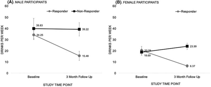
Change in drinks per week consumed through study participation. Panel A highlights drinks per week in male participants, Panel B highlights drinks per week in female participants. Reduction in drinks per week from baseline to 3-month time points in responders was statistically significant p < 0.05. Credit: Alcoholism: Clinical and Experimental Research (2023). DOI: 10.1111/acer.15123 Brain imaging of neuron activity in certain areas of the brain may predict whether an individual is likely to successfully respond to interventions to reduce their drinking.
In a study published in Alcohol: Clinical and Experimental Research , individuals whose baseline imaging showed decreased activity in areas of the brain associated with reward processing and impulsivity and increased activity in regions responsible for complex cognitive processes and emotional regulation were more likely to reduce their drinking following an intervention.
The study examined differences in pre-intervention resting state functional connectivity (rsFC) between people with alcohol use disorder who reduced their alcohol consumption and those who did not. Resting-state functional connectivity is an indirect measure of neuron activity between areas of the brain while the brain is not engaged in specific cognitive, emotional, or other tasks.
Functional Magnetic Resonance Imaging (MRI) showed significant differences in baseline resting state functional connectivity in regions of the brain associated with reward processing, stimulus prioritization, and complex problem-solving in those who responded well to interventions compared to those who did not.
People with alcohol use disorder who reduced their drinking after the intervention also showed decreased activity in the regions associated with impulsivity and alcohol valuation and increased activity in the regions responsible for higher-order cognitive processes and emotional regulation.
The patterns suggest that individuals who responded well to the intervention may have greater internal motivation or less severe disruption to the brain from their alcohol use disorder which may allow them to better engage in the intervention to reduce alcohol use and increase positive behaviors.
The Canadian study conducted functional MRIs on 46 adults with alcohol use disorder aged 21 to 55 immediately followed by a brief, approximately one-hour-long intervention aimed at reducing their alcohol use, with the option of 3 additional visits if participants chose. Three months later, researchers assessed changes in participants’ alcohol use.
Both male and female participants who responded to the intervention reduced the amount they drank per week, from 34 drinks per week at baseline to 15 at three months for male participants and 20 drinks per week to six for female participants. For those who did not respond to the intervention, the weekly number of drinks remained relatively flat for men and increased for women from 19 drinks per week at baseline to 24 drinks per week after 3 months.
The authors note that while the resting state functional connectivity patterns identified in the study may contribute to positive health behavior, the analysis is exploratory and further investigation is needed.
Provided by Research Society on Alcoholism
‘Hyper-synchronized’ brain waves may explain why different psychedelics have similar effects, rat study reveals

Psychedelics can vary by the type of receptor they target in the brain, but how does this translate to the shared hallucinogenic experiences users have? (Image credit: Pär Halje) The psychedelics LSD and ketamine work differently but induce some of the same trippy effects. A new study in rats hints at why: Both drugs trigger the same, simultaneous “wave” of electrical activity across different regions of the brain.
Psychedelic drugs are a group of substances that stimulate changes in people’s perception and cognition, sometimes referred to as a “trip.” These drugs vary in how they interact with brain cells, yet they all induce similar hallucinogenic effects — distorting users’ sense of time or causing them to perceive colors as more vivid, for example.
These overlapping effects have long puzzled scientists. But in the new rat study, published July 26 in the journal Communications Biology , scientists discovered that the reason may be that different psychedelics induce similar patterns of electrical activity in multiple regions of the brain.
“It seems to be this distinctive wave phenomenon — how the neurons behave collectively — that is most strongly linked to the psychedelic experience,” lead study author Pär Halje , a researcher in neurophysiology at Lund University in Sweden, said in a statement . Meanwhile, if you zoom in closer, the activity of individual brain cells looks quite different under the influence of ketamine or LSD, he said.
Related: Psychedelics may treat depression by invading brain cells
Psychedelic drugs are classified by the type of receptor they stimulate in the brain. “Classical” psychedelics — which include lysergic acid diethylamide (LSD), N,N-Dimethyltryptamine (DMT) and psilocybin — target 5-HT2A receptors that respond to the so-called feel-good chemical serotonin . Others, like ketamine or phencyclidine (PCP), are known as “dissociatives” and block the NMDA receptor that is usually bound by glutamate, a chemical messenger that “excites” brain cells, or pushes them toward firing.
The cellular effects of both classical and dissociative psychedelics have immediate downstream consequences that lead to the hallucinogenic symptoms that make these drugs famous.
To better understand how psychedelics work on a larger scale, Halje’s team measured variations in electrical activity in 128 regions of the rat brain, simultaneously. The rats had been given either 5-HT2A-receptor psychedelics, including LSD, or NMDA-receptor psychedelics, such as ketamine.
Both classes of drug altered the activity of two types of cell: interneurons, which connect nerve cells, and excitatory cells known as pyramidal cells . However, while LSD turned down the activity of both of these cell types, ketamine turned down only pyramidal cells and actually turned up interneurons.
Nonetheless, across many regions of the brain, both drugs triggered a similar pattern of high-frequency brain waves that unfolded at the same time, the team observed. They saw these synchronized waves in the ventral striatum, a key part of the brain’s reward system , as well as in three regions of the cortex, the outer surface of the brain.
“One might think that a strong wave starts somewhere, which then spreads to other parts of the brain,” Halje said. “But instead, we see that the neurons’ activity synchronises itself in a special way — the waves in the brain go up and down essentially simultaneously in all parts of the brain where we are able to take measurements.”
“Likely, this hyper-synchrony has major effects on the integration of information across neuronal systems and we propose that it is a key contributor to changes in perception and cognition during psychedelic drug use,” the authors wrote in the paper. In other words, neuronal information that would otherwise be isolated could be passing between different parts of the brain, leading to an altered state of consciousness.
RELATED STORIES
— Psychedelic psilocybin could treat anorexia in some patients, trial suggests
— Australia clears legal use of MDMA and psilocybin to treat PTSD and depression
The team looked only at rats, but they suggested that the study could act as a framework for future research into psychosis , during which a person loses contact with reality. “Potentially, similar mechanisms could induce hallucinations and delusions in psychotic disorders, which include and would constitute promising targets for new antipsychotic treatments,” they concluded. Such disorders include conditions like schizophrenia.
According to Halje, modeling the effects of psychedelics could even help improve our understanding of consciousness itself.
“This is where it is exciting,” he said. “The synchronized pattern we see, and whether this can help us to track down the neural foundations of consciousness.” Live Science newsletter
Stay up to date on the latest science news by signing up for our Essentials newsletter.
Contact me with news and offers from other Future brandsReceive email from us on behalf of our trusted partners or sponsorsBy submitting your information you agree to the Terms & Conditions and Privacy Policy and are aged 16 or over.
Emily Cooke
Emily is a health news writer based in London, United Kingdom. She holds a bachelor’s degree in biology from Durham University and a master’s degree in clinical and therapeutic neuroscience from Oxford University. She has worked in science communication, medical writing and as a local news reporter while undertaking journalism training. In 2018, she was named one of MHP Communications’ 30 journalists to watch under 30. ( emily.cooke@futurenet.com )
Your Pupils Can Reveal a Hidden Signal About Your Brain Function

The way your pupils react to light exercise could reveal whether you’re getting one of the key benefits of movement – the cognitive boost linked to improved mood and enhanced executive function.
Researchers in Japan monitored pupil size in 24 participants during 10 minutes of light exercise and then used neuroimaging to see how participants’ brains responded to a cognitive task.
It was only a small trial, but the results showed that the more people’s pupils dilated during light exercise – in other words, the bigger they got – the better the cognitive boost they received.
“This finding supports our hypothesis that the pupil-linked mechanisms … are a potential mechanism by which very light exercise enhances prefrontal cortex activation and executive function,” the team of neuroscientists and exercise scientists from the University of Tsukuba explain in their paper .
While it sounds like a weird link, it’s now well established that exercise, even low-intensity exercise such as yoga and walking , can boost our mood and help us focus better to complete a task – benefits that involve the brain’s prefrontal cortex .
But the exact neural activity that leads to this executive function boost from exercise is still poorly understood.
One way to get an insight into what’s going on inside our brain is through the eyes; previous research has shown that our pupils can reflect deeper neural activity.
To investigate further, the researchers recruited healthy young adults and asked half of them to participate in 10 minutes of very light exercise, and the other half to serve as a control group and simply rest on the exercise machine.
Before, during, and after the workout period, participants had their pupil size non-invasively monitored, and they were also asked about their mood.
Both groups took an executive function test before and after the workout. This was called the Stroop color task and it asked whether a word (red or green, for example) matched the color it was displayed in.
While this test was carried out, the team studied participants’ prefrontal brain activity using functional near-infrared spectroscopy.
The results showed that the light exercise made participants perform better on the cognitive task than the control group, and this was correlated with increased activity in the left dorsolateral prefrontal cortex of the brain, a region associated with executive function.
Interestingly, they also showed that the pupils of the exercise group dilated as they worked out, while the pupils of those in the control group didn’t change size.
And the more a participant’s pupils dilated during the workout, the better the boost in cognitive function when they performed the test at the end.
“These results strongly suggest that the enhancement of prefrontal executive function resulting from very light exercise can be attributed to pupil-linked neural activity,” a press release explains .
“Looking ahead, pupil diameter holds promising potential as a novel biomarker that can be used to predict the effects of exercise on the brain.”
This is the first study to suggest that the part of the central nervous system that underlies pupil dilation is linked to the cognitive boost that comes from exercise.
Of course, it’s just a small, preliminary study. The researchers explain that the results are limited by only looking at healthy young adults and an imbalanced group of men and women.
Follow-up studies are required to make sure this effect is real. And then we can start to tease out what it means, and how we can benefit from it.
But it’s a fascinating new insight into the link between our brains and our eyes – as well as the way exercise can alter our brain function.
The research has been published in NeuroImage .
Memory Expert Reveals Why Reading Books Can Improve Your Brain Health
Whether you’re into biographies or bodice-ripping romances, there’s nothing like a good book. Since the first novel was published in Japan 1,000 years ago, we’ve been transported by the power of words, but reading doesn’t just boost your imagination—it can also strengthen your brain.
“Reading activates neural circuits controlling vision, language, and learning and can lead to improved memory, thinking and mood,” Dr. Gary Small, a geriatric psychiatrist and one of America’s leading memory experts, told Newsweek .
But you don’t have to power through a 500-page paperback in one sitting to reap the benefits. Here’s why reading a book is good for you, and how you can fit this brain-booster into daily life. A stock image of a young woman carrying a stack of books. Reading doesn’t just boost your imagination—it can also strengthen your brain. iStock / Getty Images SUBSCRIBE NOW FROM JUST $1 > Why is Reading Good For Your Brain?
Reading is actually a pretty impressive skill and one that sets us apart from other species in the animal kingdom. Research by MIT suggests that our brains “recycled” part of our visual cortex, the area responsible for recognizing objects, to understand patterns, allowing us to read. So, it’s no surprise that books have a noticeable impact on our noggins.
“We can measure these effects with brain scans and neuropsychological tests that assess cognitive abilities and mood states,” Small said. “Reading will stimulate the brain’s left hemisphere in temporal, frontal and parietal regions that control language, speech, thinking and mood.”
Here are five reasons why reading is good for your brain.
SIGN UP FOR NEWSWEEK’S EMAIL UPDATES > It Can Improve Your Concentration
Struggling to focus? According to Small, reading strengthens the white matter in your brain, making it easier to concentrate.
“White matter functions as insulation, wrapping around long nerve cell wires,” Small said. “These wires serve as communication connections between brain cells.”
White matter helps to relay information between different areas of your brain and it is important for focus and learning. A 2012 study by the University of Stanford found that the white matter of children with below-average reading skills increased when they read more, with the effects measured over a three-year period. A stock photo of a young woman reading a book. Reading strengthens the white matter in your brain, making it easier to concentrate. Daniel de la Hoz/iStock/Getty Images Plus It Can Boost Your Mood and Reduce Anxiety
If you’re having a bad day, swap the doom-scrolling for a book. While endlessly thumbing through social media can give you a quick dopamine hit, it can also leave you feeling low. However, literature, particularly fiction, can improve your mood .
“Reading can reduce anxiety,” Small said. “Becoming engrossed in a compelling narrative helps anxious people escape their obsessive and negative thinking.” It Can Spark Your Creative Instincts
If you find yourself stuck in a creative rut, then reading might be the answer to your imagination’s prayers. A study published in the journal Thinking Skills and Creativity surveyed almost 200 university students and found that those that read or write regularly scored higher in a creativity test than those who did not.
“When we read, we use our imagination, which strengthens the neural circuits controlling that mental task,” Small said. “Considering the ideas and creativity of the material sparks the reader’s ability to come up with novel thoughts.” It Can Boost Your Vocabulary
With research showing that vocabulary is important for academic and social opportunities, expanding your lexicon can only be a good thing. Fortunately, reading regularly means you’re less likely to be lost for words.
“When we read, we learn new words and concepts that enhance vocabulary and creative thinking,” Small said.
But put that dictionary down, as reading something you love is just as beneficial, according to Small.
“Reading is an enjoyable form of brain exercise that helps us relax, escape our everyday worries, and reduce stress levels,” he said. “Less stress translates to less distraction and better cognitive performance.” A stock photo of a grandfather reading a book to his grandson. Reading has been proven to boost brain power for all ages. evgenyatamanenko/iStock/Getty Images Plus It Can Slow Age-Related Mental Decline
Although aging is inevitable, there are things you can do to keep your brain sharper for longer—and reading is one of them.
In a 14-year study published in the journal International Psychogeriatrics , researchers tracked the mental decline of 1,900 people aged 64 and over. They found that people who read more than once a week retained cognitive function better than non-readers, regardless of their education level.
“Many stimulating mental activities, such as reading, have been linked to slower cognitive decline,” Small said. “Mental stimulation activates neural circuits, which strengthens and protects them from age-related dysfunction.” Should I Read Fiction or Non-Fiction?
Although both fiction and non-fiction are good for your brain health, they have different advantages.
“A 2013 study published in Science showed that literary fiction can improve the ability to interpret another person’s mental state, intentions and beliefs,” Small said. “Reading non-fiction is thought to improve concentration, general knowledge and problem-solving skills.” What’s the Difference Between a Paper Book and a Screen?
A 2018 study involving more than 170,000 readers and published in the Educational Research Review discovered that reading comprehension was better for book lovers than screen fans.
Reading online comes with more distractions than reading from a page, meaning you’re more likely to skim the content and retain less information.
But what about e-readers? Experts are split on which category devices such as Amazon Kindles fall into, but they do emit blue light, which can interfere with your sleep in the same way cell phones and TV screens do. A stock photo shows a man in a library. Reading both fiction and non-fiction is good for your brain. anyaberkut/iStock/Getty Images Plus How Often Should I Read to See Benefits? If you’ve never been much of reader or you’ve fallen out of the habit, Small recommends starting slowly.”My studies of brain function and health suggest that it’s best to train but […]
A new mechanism encouraging the brain to self-repair after an ischemic stroke
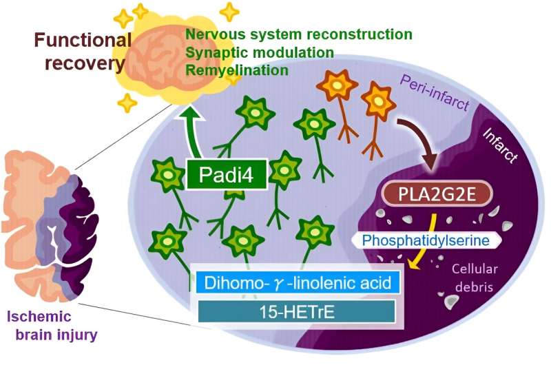
The neurons around an injured brain region will play major roles in promoting functional recovery through nervous system development, synapse organization, and remyelination; however, the molecular mechanisms triggering such a broad range of neural repair after brain injury remained unknown. Our study demonstrated that the secretion of PLA2G2E from perilesional neurons with ischemic stress generated DGLA and 15-HETrE, which triggered these recovery processes through the neuronal Padi4-dependent induction of cluster formation associated with neural repair after ischemic brain injury. Credit: Department of Neuroinflammation and Repair, TMDU Patients often experience functional decline after an ischemic stroke, especially due to the brain’s resistance to regenerate after damage. Yet, there is still potential for recovery as surviving neurons can activate repair mechanisms to limit and even reverse the damage caused by the stroke. How is it triggered though?
In a study published recently in Neuron , researchers from Tokyo Medical and Dental University (TMDU) provided new insights regarding this question by identifying a new mechanism. They discovered that neurons surrounding the area of cell death secrete lipids that can trigger brain -autonomous neural repair after ischemic brain injury.
An ischemic stroke occurs when the blood supply to the brain is blocked and results in the death of brain cells. This condition is life-threatening, and patients will likely develop functional disabilities. Although the adult brain can self-repair, the underlying mechanisms need further clarification.
Inflammation of the brain contributes to the effects of ischemic stroke . “There is evidence that more lipids are produced after tissue injuries and contribute to regulating inflammation,” says Takashi Shichita, senior author of the study. “We investigated the changes in lipid metabolite production in mice after ischemic stroke. Interestingly, the levels of a specific fatty acid called dihomo-γ-linolenic acid (DGLA) and its derivatives increased after the stroke.”
The researchers further discovered that a protein known as PLA2GE2 (Phospholipase A2 Group IIE, an enzyme) mediates DGLA increase. By manipulating the expression of PLA2GE2, they also showed its impact on functional recovery. Deficiency of PLA2GE2 led to more inflammation, lower expression of factors stimulating neuronal repair, and more tissue loss. The team carried on with identifying the targets of PLA2GE2/DGLA.
“When we look at genes expressed in mice lacking PLA2GE2, we found low levels of a protein called peptidyl arginine deiminase 4 (PADI4),” explains Akari Nakamura, lead author of the study. “PADI4 regulates transcription and inflammation. Remarkably, expressing PADI4 in mice limited the extent of tissue damage and inflammation after ischemic stroke.”
Additionally, the study shows that PADI4 promotes the transcription of genes involved in brain repair. It also identifies the whole signaling pathway involved in this process.
Most data were obtained in a mouse model of ischemic stroke. Yet, the recovery pathway likely exists in humans as the researchers found that neurons surrounding the stroke site express PLA2G2E and PADI4 in humans. Moreover, another recent study reported that the lower serum DGLA level was correlated with the severe ischemic stroke and cognitive disorders in humans.
This study describes a new mechanism that triggers brain repair after an ischemic stroke, which might lead to the development of compounds promoting PADI4’s effects, that stimulate the recovery of patients. It could also change our current understanding and approach toward Eicosapentaenoic acid (EPA) or Docosahexaenoic acid (DHA), as the only beneficial lipids for preventing atherosclerosis and vascular diseases.
Provided by Tokyo Medical and Dental University
Sleep deprivation linked to health issues like obesity and cognitive decline

Decentralize.TV – Episode 9 – Aug 8, 2023 – FIRO innovator Reuben Yap on PRIVATE digital money Advertisement
It’s normal to sometimes have trouble falling asleep at night, but if you can’t sleep well three days a week or your sleep problems persist for one month or longer, you may have sleep disorders that require immediate attention .
An estimated 50 to 70 million people in the U.S. suffer from chronic or ongoing sleep disorders, according to the National Heart, Lung and Blood Institute (NHLBI).
If you think you may have the same problem, you can improve your sleep quality by making some lifestyle changes. Major types of sleep disorders
Here are four common types of sleep disorders .
Difficulty falling asleep
One of the most common issues faced by people with sleep disorders is difficulty falling asleep. Ideally, children or adolescents should fall asleep within 30 minutes of lying in bed with their eyes closed.
Meanwhile, adults should fall asleep within one hour under the same conditions. (Related: State of insomnia: Study ranks California as the No. 1 most sleep-deprived state .)
Shallow sleep
Some people with sleep disorders can fall asleep, but their problem is that they often experience shorter periods of deep sleep and spend more time in a lighter sleep state, which still causes poor sleep quality.
Even if they get at least seven to eight hours of sleep , these people usually wake up feeling unrefreshed and fatigued.
Other people with health issues like allergies, frequent nasal congestion or thick nasal polyps may have to breathe through their mouths, resulting in a dry mouth and shallow sleep.
While individuals with sleep apnea may not always have nasal congestion, they still need to breathe through their mouths due to nighttime breathing interruptions.
And if people with this sleep problem experience a particularly dry mouth during the night, they may wake up for water or feel very thirsty in the morning. They may still feel unrefreshed even after sleeping for a long time.
According to Traditional Chinese Medicine (TCM), meridians are the channels where life energy (qi) flows in the body. Meridians are responsible for transporting qi and blood throughout the entire body, which are the crucial substances that constitute and sustain human life.
The body consists of 12 major meridians, each corresponding to a specific organ. The internal organs are connected to the body’s surface through these meridians.
The meridians house specific points called acupoints with unique functions. By stimulating the corresponding acupoints through techniques like acupuncture and massage , you may relieve health issues linked to specific organs.
Nighttime awakenings
People with this sleep disorder often experience multiple awakenings during the night.
This can be a problem because every time they wake up, it takes them more than 30 minutes to fall back asleep.
Early morning a wakening
People suffering from early morning awakenings usually wake up more than an hour earlier than their usual time. Additionally, their overall sleep duration during the night is often less than five hours.
This can cause issues because the recommended daily sleep duration is at least 6.5 to eight hours.
If you don’t have trouble falling asleep at night, you should also make sure you don’t oversleep because it can also make you feel more tired.
TCM practitioners say that “prolonged bed rest weakens qi,” meaning that extended periods of lying down in bed can hinder the circulation of qi and blood in your body.
To maintain your overall health, it is better to avoid extended bed rest even if you are experiencing discomfort or pain. Experts say engaging in appropriate physical activity may offer more benefits than longer bedrest for overall health. Health issues linked to poor sleep One of the most direct impacts of sleep problems is fatigue caused by insufficient sleep.Sleep disorders can also have these negative side effects: Cognitive impairment Data suggests that inadequate sleep can result in a decline in brain function , including memory.The hippocampus, located on the sides of your brain, has an important role in storing short-term memories. Throughout the day, your memories are stored in the frontal lobe, but during nighttime sleep, they are transferred to the hippocampus and are then transferred back to the frontal lobe for permanent storage.Poor sleep can impact the functionality of the hippocampus, meaning it can affect the consolidation of temporary memories into permanent ones. Increased risk of dementia Many elderly dementia patients have chronic sleep disorders because not getting enough sleep can impact memory storage and prevent the brain from resting properly.Chronic poor sleep may also contribute to brain atrophy and exacerbate the degeneration of brain function. Increased obesity risk Your body’s metabolism functions better with adequate sleep, while poor sleep can contribute to weight gain.This means getting enough quality sleep is essential, especially if you want to lose weight or sustain a healthy weight.If you have high blood lipids even after taking cholesterol-lowering medications or following a balanced diet, you may want to consult your doctor to check for sleep disorders.Improving sleep quality can boost metabolism, which may help address any lipid metabolism disorders. Metabolic syndrome Chronic sleep disorders can cause metabolic disorders such as high blood sugar, high blood lipids, hypertension and obesity.In severe cases, it can even increase the risk of cardiovascular diseases, including heart disease and stroke. Weakened immune system Insufficient sleep can disrupt the body’s hormones, endocrine system and nervous system, which may contribute to a weakened immune system. Natural and drug-free ways to improve sleep quality Try the tips below if you have trouble falling asleep every night: Sunbathe Sunbathing can regulate your autonomic nervous system and melatonin secretion, which can help improve sleep quality at night.Try to spend at least 30 minutes sunbathing in the morning or go for a run or another activity for one hour during sunset. Even on cloudy or rainy days, the sun’s photons can still reach the Earth and offer benefits, making outdoor activities conducive to your well-being. Foot soak Try soaking your feet in hot water before going to bed. The closer the […]

The Power Within by Corey Daniels book available for only $2.99
Mind has both conscious and subconscious halves. These are likened to a driver and the truck he drives. The driver plans the destination and observes road conditions, while the truck provides motive power. Your subconscious mind is like the truck and it only goes in the direction in which is a steered. This can be the road or off a cliff. Likewise, the consciousness paints a picture of what the world is and what your goals are and the subconscious acts on them through emotion, physical response and energy, whether these are correct, rational images or false negative ones. The subconscious is also like an emotional reservoir which your body and mind draw responses from to external stimuli.
The Power Within lays out a method of programming your subconscious and tapping into the Holy Spirit, God voice or what the Greeks called the daimon (godman).