Nature Knows and Psionic Success
God provides
Brain networks encoding memory come together via electric fields, study finds
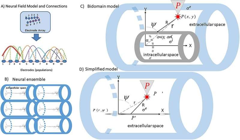
A) Neural field model and connections. Neural fields provided a quantitative way to describe each ensemble’s patterns of activity across simultaneously recorded sites. The same model can describe different ensembles. Each electrode occupies a position on a cortical manifold (line) Δ parameterized by the variable x and is connected to all other electrodes with connections whose strength follows a Gaussian profile (colored solid and dashed lines), see (Pinotsis et al. 2017) for more details. B) Extracellular space around each neuron within the ensemble (blue cylindrical fibers). C) Bidomain model for the electric field generated by a cylindrical fiber in a conductor. The extracellular and intracellular space are depicted by blue and gray cylindrical fibers (see Methods for the meaning of various symbols). D) Simplified bidomain model where the measurement point is located at a vertical distance much larger than the radius of intracellular space. Credit: Cerebral Cortex (2023). DOI: 10.1093/cercor/bhad251 The “circuit” metaphor of the brain is as indisputable as it is familiar: Neurons forge direct physical connections to create functional networks, for instance to store memories or produce thoughts. But the metaphor is also incomplete. What drives these circuits and networks to come together? New evidence suggests that at least some of this coordination comes from electric fields.
The new study in Cerebral Cortex shows that as animals played working memory games, the information about what they were remembering was coordinated across two key brain regions by the electric field that emerged from the underlying electrical activity of all participating neurons. The field, in turn, appeared to drive the neural activity , or the fluctuations of voltage apparent across the cells’ membranes.
If the neurons are musicians in an orchestra, the brain regions are their sections, and the memory is the music they produce, the study’s authors said, then the electric field is the conductor.
The physical mechanism by which this prevailing electric field influences the membrane voltage of constituent neurons is called “ephaptic coupling.” Those membrane voltages are fundamental to brain activity. When they cross a threshold, neurons “spike,” sending an electrical transmission that signals other neurons across connections called synapses.
But any amount of electrical activity could contribute to a prevailing electric field which also influences the spiking, said study senior author Earl K. Miller, Picower Professor in the Department of Brain and Cognitive Sciences at MIT.
“Many cortical neurons spend a lot of time wavering on verge of spiking” Miller said. “Changes in their surrounding electric field can push them one way or another. It’s hard to imagine evolution not exploiting that.”
In particular, the new study showed that the electric fields drove the electrical activity of networks of neurons to produce a shared representation of the information stored in working memory, said lead author Dimitris Pinotsis, Associate Professor at City—University of London and a research affiliate in the Picower Institute. He noted that the findings could improve the ability of scientists and engineers to read information from the brain, which could help in the design of brain-controlled prosthetics for people with paralysis.
“Using the theory of complex systems and mathematical pen and paper calculations, we predicted that the brain’s electric fields guide neurons to produce memories,” Pinotsis said. “Our experimental data and statistical analyses support this prediction. This is an example of how mathematics and physics shed light on the brain’s fields and how they can yield insights for building brain-computer interface (BCI) devices.” Fields prevail
In a 2022 study , Miller and Pinotsis developed a biophysical model of the electric fields produced by neural electrical activity. They showed that the overall fields that emerged from groups of neurons in a brain region were more reliable and stable representations of the information animals used to play working memory games than the electrical activity of the individual neurons.
Neurons are somewhat fickle devices whose vagaries produce an information inconsistency called “representational drift.” In an opinion article earlier this year, the scientists also posited that in addition to neurons, electric fields affected the brain’s molecular infrastructure and its tuning so that the brain processes information efficiently.
In the new study, Pinotsis and Miller extended their inquiry to asking whether ephaptic coupling spreads the governing electric field across multiple brain regions to form a memory network, or “engram.”
They therefore broadened their analyses to look at two regions in the brain: The frontal eye fields (FEF) and the supplementary eye fields (SEF). These two regions, which govern voluntary movement of the eyes, were relevant to the working memory game the animals were playing because in each round the animals would see an image on a screen positioned at some angle around the center (like the numbers on a clock). After a brief delay, they had to glance in the same direction that the object had just been in.
As the animals played, the scientists recorded the local field potentials (LFPs, a measure of local electrical activity) produced by scores of neurons in each region. The scientists fed this recorded LFP data into mathematical models that predicted individual neural activity and the overall electric fields.
The models allowed Pinotsis and Miller to then calculate whether changes in the fields predicted changes in the membrane voltages, or whether changes in that activity predicted changes in the fields. To do this analysis, they used a mathematical method called Granger Causality.
Unambiguously this analysis showed that in each region, the fields had strong causal influence over the neural activity and not the other way around. Consistent with last year’s study, the analysis also showed that measures of the strength of influence remained much steadier for the fields than for the neural activity, indicating that fields were more reliable.
The researchers then checked causality between the two brain regions and found that electric fields, but not neural activity, reliably represented the transfer of information between FEF and SEF. More specifically, they found that the transfer typically flowed from FEF to SEF, which agrees with prior studies of how the two regions interact. FEF tends to lead the way in initiating an eye movement.
Finally, Pinotsis and Miller used […]
Read more at medicalxpress.com

The Power Within by Corey Daniels book available for only $2.99
Mind has both conscious and subconscious halves. These are likened to a driver and the truck he drives. The driver plans the destination and observes road conditions, while the truck provides motive power. Your subconscious mind is like the truck and it only goes in the direction in which is a steered. This can be the road or off a cliff. Likewise, the consciousness paints a picture of what the world is and what your goals are and the subconscious acts on them through emotion, physical response and energy, whether these are correct, rational images or false negative ones. The subconscious is also like an emotional reservoir which your body and mind draw responses from to external stimuli.
The Power Within lays out a method of programming your subconscious and tapping into the Holy Spirit, God voice or what the Greeks called the daimon (godman).