Nature Knows and Psionic Success
God provides
The Three Channels of Gut-Brain Communication

Key points
Gut microbes communicate with the brain using three different channels.
The nervous system is the fastest of the three.
The immune system is potent, but lacks subtlety.
Hormones are slow, but long acting.
Nature is everywhere gothic, not classic. She forms a real jungle, where all things are provisional, half-fitted to each other, and untidy.— William James New discoveries about the gut-brain axis continue to illuminate how gut microbes affect our mood. Even after years of working on the concept, scientists are still amazed at how tiny microbes can alter the workings of the mighty human brain.
Since we can’t escape the dangerous germs coating our planet, animals have adopted their own set of beneficial microbes to fight, starve, and outcompete the pathogens. Like all important biological functions, there are backups and backups to the backups. And so, for our friendly microbes, our body has created at least three different avenues of communication. Three different approaches to the brain. Courtesy of Midjourney
Each communication channel has its charms. Some are fast and potent, while others are gentle and languorous. If you’ve ever had food poisoning, you know about fast and potent. When your gut senses an attack of nasty microbes, you receive an urgent directive to find a bathroom, pronto. On the other hand, when you enjoy a warm bowl of oatmeal, your mood may reflect the laid-back consensus of contented gut microbes.
The three main communication channels between your brain and your gut include your nervous system , your immune system, and your endocrine system. The circulatory and lymphatic systems also play supporting roles, but these first three are dominant. Nevertheless, each system interacts with the others, making it difficult to discuss any of them in isolation. William James was right; nature is untidy.
Some of the following includes excerpts from my book The Psychobiotic Revolution that I wrote with researchers Ted Dinan and John Cryan of University College Cork, in Ireland. They are major players in the gut-brain field, and they coined the term psychobiotic to refer to microbes that can improve mood. If you are considering a deep dive into the science of the gut-brain axis, they are essential reading.
Each of the gut-brain channels has its own unique chemical language, but because they also communicate with each other, they need to have some signaling molecules in common. Here are the three main systems and the chemicals they use to chat. The nervous system
The nervous system relays information to and throughout your brain. It communicates using chemicals called neurotransmitters. Its communication style is fast and point-to-point, but short-acting. The vagus nerve is the primary boulevard for this neural traffic.
article continues after advertisement
Your gut is encased in a tube of nerves called the enteric nervous system. It contains as many nerve cells as your spinal cord, and in honor of its size and importance, it is called the second brain. A big part of its job is to control peristalsis, the rhythmic movement of muscles that squeeze your food from one end to the other. But your gut lining also contains cells that can sense the environment and send messages to the brain. These can be notices of satiety or more ominously, about pathogens and food poisoning.
Incredibly, the microbes in your gut produce neurotransmitters identical to those that your brain uses to communicate from one cell to another. These are partly for microbes to talk to each other, but also to talk to their host. The neurotransmitters include dopamine and serotonin, two of the most important chemicals involved in psychiatry . This is thus an immediate channel of communication for your gut microbes to let your brain know what’s going on down under. It can quickly alter your mood.
This gut-brain connection makes it clear why you should be solicitous of your microbial happiness . Treat your microbes right, and they will be on the front line of defense against pathogens. Abuse them, and they can make you miserable on short notice. The immune system
The immune system is always primed to rally a defense against pathogens. It uses proteins called cytokines to signal distress. It can communicate quickly, but its urgent chemical effect can be harsh enough to cause tissue damage.
In the first 1,000 days of your life, your immune system is tasked with learning to accommodate your good gut bacteria while still remaining vigilant against pathogens. Immune cells called regulatory T cells can recognize beneficial bacteria and tamp down any attempt to destroy them. After that early education , your immune system settles down to a lifetime of fighting black-hatted germs while protecting your white-hatted good gut microbes.
However, this is situational. Beneficial microbes in your gut are tolerated, but if they breach the gut lining and gain entry into the bloodstream, your immune system will track them down and kill them. Your brain is kept in the loop on these systemic infections and may exhibit sickness behavior, which is similar to depression : you just want to be left alone to let your body heal.
article continues after advertisement
Your brain is protected from blood-borne microbes by the blood-brain barrier. But if a systemic infection rages on without resolution, that barrier can weaken. If toxins or bacteria enter the brain, anxiety or even psychopathy can result. Your immune system will chase these rogue microbes into the brain, killing them but also causing collateral damage. In this scenario, your immune system can do more damage than the microbes that stimulated it. The endocrine system
The endocrine system uses hormones to monitor and manage growth and metabolism. Its component glands communicate by secreting these hormones into your blood and thus send signals throughout the body. Its operations are slower, more moderate, and systemic, but longer acting than those of the other two systems.
The most pertinent of the endocrine systems connecting the gut and the brain is the hypothalamus, pituitary, and adrenal trio – the so-called HPA axis. The hypothalamus is always testing the blood and when it senses stress , it signals […]
How a sunny outlook toward ageing can help to protect your memory

Pic: iStock A POSITIVE attitude towards ageing can help you ward off mild cognitive impairment better than people who become grumpier and more cantankerous. It certainly seems to be working for 96-year-old David Attenborough, who said last year that “focus and curiosity” help him stay clear-minded. And researchers at Yale University School of Public Health have shown how a positive attitude could help the rest of us avoid or even reverse age-related memory loss and restore cognitive ability to normal.
Forgetting names of people and places and appointments and misplacing things are all common symptoms of age-related MCI (mild cognitive impairment), which, according to the Irish Longitudinal Study on Ageing, affects as many as 10% of people in Ireland. “Most people assume there is no recovery from MCI, but, in fact, half of those who have it do recover,” says Becca Levy, public health and psychology professor at Yale and lead author of the new study. “Little is known about why some recover while others don’t.”
To determine if an optimistic outlook on ageing has any effect, Prof Levy and her team recruited 1,716 people in their 70s enrolled in the US National Health and Retirement Study, some of whom were diagnosed with MCI and others who had normal cognition. Participants were tracked for 12 years, and results published in JAMA Network Open journal showed that those with a cheery disposition had a 30.2% greater likelihood of recovering from memory impairment than those with negative age beliefs.
A positive outlook also meant people recovered their cognition up to two years faster than the grumpy cohort. Prof Levy said that “age-belief interventions could increase the number of people who experience cognitive recovery”.
The new findings add to a slew of evidence about the power of positivity on healthy ageing for mind and body. Three years ago, psychologists from Northwestern University reported that, of 991 middle-aged and older people, those who were mostly enthusiastic about the future performed better in memory tests as they got older. Others have shown that optimistic adults have better blood sugar control and healthier cholesterol levels, adding up to improved cardiovascular health, and that a habitually positive outlook on life can minimise chronic pain and associated emotional distress.
Another recent study of 1,000 younger adults in Europe, Canada, and the US revealed that dark feelings are more likely linked with poor health, including stress-related headaches, nausea, back pain, and insomnia. Professor Reinhard Pekrun, a psychologist at the University of Essex who led the investigation, says his findings suggest that if two people of equal cognitive ability took a test, the more positive-minded individual would achieve a grade higher than someone with a negative mindset. “Overall, hope was the healthiest and best way to spark success and promote long-term happiness,” Prof Pekrun says.
If you are prone to pessimism, you will also be accumulating stress. “Over time, being negative and self-critical means your body will begin to shut down to protect itself, becoming slower and more susceptible to health conditions, such as depression, anxiety, and sleep problems,” says psychologist Dr Julie Hannan, author of The Mid Life Crisis Handbook.
The good news is that you can train yourself to become more positive. “Research shows that starting to give ourselves a bit of compassion and speak more kindly to ourselves is the first step out of negative thinking and away from an inner critical voice,” Dr Hannan says. “When we do this more often, the body starts to release more feelgood oxytocin and lower levels of damaging stress hormones, such as cortisol.”
Along with fostering a positive outlook, here are five steps you can take to boost your memory:
Eat plenty of fruit and veg: A diet low in beneficial plant compounds, called flavonoids, will accelerate age-related memory loss, according to a new study by researchers at Columbia and Brigham and Women’s Hospital and Harvard University. But replenishing flavonols in the diets of people over 60 produced better performances in memory tests, they found.
“The improvement among study participants with low-flavanol diets was substantial and raises the possibility of using flavanol-rich diets or supplements to improve cognitive function in older adults,” said Adam Brickman, professor of neuropsychology at Columbia University and one of the study authors.
Take a multivitamin: Another recent study of more than 3.500 adults by the same Columbia University and Brigham and Women’s Hospital team showed that popping a multivitamin supplement can slow age-related memory decline. According to JoAnn Manson, head of the Division of Preventive Medicine at Brigham and Women’s Hospital and one of the authors, “multivitamin supplementation holds promise as a safe, accessible, and affordable approach to protecting cognitive health in older adults”. However, a medical professional should always be consulted before taking any form of supplement.
Go for a daily walk: Going for a stroll will strengthen connections in and between brain networks, enhancing memory and slowing age-related cognitive decline, according to researchers at the University of Maryland. They found that older adults with MCI who walked on a treadmill four days a week during a three-month study had a slower decline in memory than non-walkers.
“These results provide even more hope that exercise may be useful as a way to prevent or help stabilise people with mild cognitive impairment and, maybe, over the long term, delay their conversion to Alzheimer’s dementia,” said Carson Smith, a professor with the School of Public Health and lead author of the study.
Drink three cups of tea a day: Tea leaves are rich in flavonoids with anti-inflammatory properties that have been shown to offer vascular protection for the brain. In 2017, research by assistant professor Feng Lei , of the National University of Singapore’s department of psychological medicine, showed that drinking at least one cup (but preferably three or more) of green or black tea a day helped to cut the risk of memory decline and dementia among older adults by 50%. Among those genetically predisposed to developing Alzheimer’s disease, the potential risk was cut by 86%.
Eat more fibre: Researchers have shown that when fibre is fermented by […]
Can Smartphone Use Increase Brain Cancer Risk?
Photo Illustration by Lecia Landis for Verywell Health; Getty Images Fact checked by Nick Blackmer Key Takeaways
People have long wondered whether cell phone use might cause brain cancer, but there’s been no evidence to prove this connection.
A new 20-year study from Taiwan aimed to answer this question, but it only added to the mountain of inconclusive research after finding a positive but ultimately weak, non-significant association between mobile phone use and brain cancer.
Experts say we should still be aware of the potential risks and take possible precautions in the meantime.
The conversation about whether radiation emitted by cell phones can cause brain cancer has been ongoing for years, though research has yet to prove a solid connection between the two.
A new 20-year study from Taiwan aims to answer this longstanding question. The population-based study examined whether smartphone usage over a 20-year period had any effect on the incidence and mortality rates of malignant brain cancers of its participants.
Researchers found a positive, but ultimately weak and non-significant association between mobile phone use and brain cancer.
“There appears to be a limited connection between mobile phone usage and MNB (malignant brain neoplasm) occurrence and mortality. However, it is crucial to recognize that definitive conclusions cannot be made at this point,” said Shabbir Syed Abdul, MD, MSc, PhD , a professor of artificial intelligence and digital health and a co-author of the study.
While the study offers some valuable insights, it doesn’t provide a concrete answer to the question at hand. It just shows that in tandem with a major increase in smartphone usage between 2000 and 2019, brain cancer incidence and mortality slightly increased as well.
The existing information on the subject is equally conflicting. In 2011, the International Agency for Research on Cancer (IARC) classified radiofrequency electromagnetic non-ionizing radiations as “possibly carcinogenic to humans,” while the 2020 guidelines drafted by the International Commission on Non-Ionizing Radiation Protection (ICNIRP) suggest that “the great majority of studies have reported a lack of carcinogenic effects in a variety of animal models.”
Related:
There’s a reason science can’t quite seem to figure out a definitive answer when it comes to phones and brain cancer, said , a neuro-oncologist at Lehigh Valley Health Network (LVHN). Morrison said measuring the impact of our phones on our health over a long period of time is tricky given how much our phone use, and the phones themselves, have changed.
MIND diet for brain health shows surprising results in new clinical trial

Editor’s Note: Sign up for CNN’s Eat, But Better: Mediterranean Style. Our eight-part guide shows you a delicious expert-backed eating lifestyle that will boost your health for life .
Results are in from the highly anticipated clinical trial on the Mediterranean-DASH Diet Intervention for Neurodegenerative Delay or MIND diet — a diet designed specifically to boost the brain — and they are less stellar than anticipated.
“We really expected that the MIND diet would show an effect above the control group, so we were quite surprised by the outcome ,” said lead study author Lisa Barnes, associate director of the Alzheimer’s Disease Research Center at Rush University Medical Center in Chicago.
Actually, the MIND diet did improve the brains of those who followed it for three years. At the end of the study magnetic resonance imaging (MRI) scans showed fewer white matter hyperintensities (tiny lesions) and a larger volume of both grey matter (the brain’s cognitive center) and white matter (the brain’s communication highway).
But here’s the rub — the brains of the control group who were not eating the MIND diet also improved to a similar degree.
Past studies have shown both the MIND diet and the Mediterranean diet significantly reduced the risk of cognitive decline and Alzheimer’s disease . However, many of the studies had a much longer duration, Barnes said.
“My main concern with this trial from the beginning has been that three years may be too short a time to have an impact on a disease process that develops over many decades,” said leading nutrition researcher Dr. Walter Willett, a professor of epidemiology and nutrition at Harvard T.H. Chan School of Public Health and professor of medicine at Harvard Medical School.
Willett pointed to an older clinical trial which found eating more beta-carotenoids, the antioxidants found in red, yellow, orange and dark green fruits and vegetables, produced cognitive benefits — but only after years on the diet.
“After 15 or more years of beta-carotene supplementation there was significant and importantly better cognitive function in the beta-carotene group compared to placebo, but after just several years there was no difference,” said Willett, who was not involved in the new study.
In addition, people in the new study’s control group may have improved their own diet instead of sticking to instructions to eat as they always had, said Barnes, who is presenting her paper Tuesday at the 2023 Alzheimer’s International Conference in Amsterdam.
“It’s not like people who were on the control diet stayed flat,” she said. “Everyone was eating healthier, losing weight, and so they all got better. My takeaway is that regardless of the type, a healthy diet does seem to improve cognitive function.”
It’s difficult to do a long-term clinical trial in nutrition because people may realize which arm of the study they are in, said Dr. David Katz, a specialist in preventive and lifestyle medicine who founded the nonprofit True Health Initiative , a global coalition of experts dedicated to evidence-based lifestyle medicine. He was not involved in the study.
“Enrolling in the study likely heighted awareness of prudent dietary practices to protect cognition among people already concerned about that,” Katz said. “This study did not exclude a difference; it simply failed to confirm one.” What is the MIND diet?
Developed in 2015 by researchers at Rush University in Chicago, the MIND diet incorporates much of the plant-based Mediterranean diet , which focuses on fruits, vegetables, whole grains, beans, seeds, nuts and a lot of extra-virgin olive oil. Red meat and sweets are eaten rarely, but fish, which are packed with good-for-you omega-3 fatty acids, are a staple.
The MIND diet also assimilates elements of the Dietary Approaches to Stop Hypertension (or DASH) diet. The DASH diet focuses on lowering blood pressure and cholesterol which can lead to heart attacks, strokes, and constriction of small blood vessels that can lead to dementia. The standard DASH diet limits salt to 2,300 milligrams a day, less than a teaspoon of table salt. Despite the disappointing results of this trial, other studies have found that the MIND diet can reduce the risk of cognitive decline and Alzheimer’s disease. – vaaseenaa/iStockphoto/Getty Images Numerous studies have found the Mediterranean diet can reduce the risk for diabetes , high cholesterol , dementia , memory loss , depression and breast cancer . The diet, which is more of an eating style than a restricted diet, has also been linked to stronger bones , a healthier heart and longer life . The DASH diet has been shown to reduce blood pressure and is the American Heart Association’s top diet.
The MIND diet takes the Mediterranean and DASH diets to the next level by focusing on foods known to boost brain health. Dark leafy greens should be eaten every day of the week on the MIND diet. Those include arugula, collards, dandelion greens, endive, grape leaves, kale, mustard greens, romaine lettuce, spinach, Swiss chard and turnip greens.
Berries are also stressed over other fruits on the MIND diet. Blackberries, blueberries, raspberries or strawberries should be eaten at least five days a week.
In addition, three servings of whole grain should be eaten daily. Beans should be eaten in four meals a week, poultry in two and fish at least once a week. Eat nuts five times a week, and avoid butter, cheese, red meat, fried foods and pastries and sweets.
A 2017 study of nearly 6,000 healthy older Americans with an average age of 68 found those who followed the Mediterranean or MIND diet lowered their risk of dementia by one-third. Both groups lost weight
The study, published Tuesday in the journal New England Journal of Medicine, followed 604 overweight people over 65 for three years. All had a first-degree relative with Alzheimer’s disease and were cognitively normal at the start of the study.
The experimental group was asked to follow the MIND diet while also cutting 250 calories a day with the help of a counselor. Vitamin supplements were not allowed. This group was provided with appropriate amounts of olive oil, blueberries and nuts each month.
The control […]
The #1 Brain Exercise for Memory Improvement, According to Neurologists

You likely already know to brush teeth to prevent dental cavities, work out to s trengthen muscles and wear sunscreen to protect skin, you may not realize there are things you can do to keep your brain sharp .
For starters, a 2019 study of nearly 200,000 adults found that those who had a healthier lifestyle were less likely to develop dementia over the course of eight years, even if they were genetically at risk for dementia — and a 2020 study came to a similar conclusion. Beyond general healthy habits, though, specific activities have been shown to boost brainpower and prevent cognitive decline: brain exercises. What are brain exercises?
Simply put, brain exercises are activities that engage your brain. They include everything from computer-based puzzle games and reading to playing sports and talking to people. The key with brain exercises is that to really be effective, they have to require you to actively participate in them. Things that are more passive, such as watching TV, give you some visual stimulation, but there’s no back-and-forth engagement, explains Douglas Scharre, M.D. , the director of The Ohio State Wexner Medical Center Division of Cognitive Neurology. “That’s probably less a benefit to your brain than activities that you’re more involved in.” Do brain exercises work?
Probably — but it’s complicated. “Memory is not one thing, but it’s a combination of different things so when we talk about exercises or training for memory, I think it depends on what type of memory we’re referring to,” says Zaldy S. Tan, M.D., M.P.H. , the director of the Cedars-Sinai Health System Memory and Aging Program. Consider a trip to the grocery store: Remembering what you intended to buy without having a list to look at requires an ability to recall things. Remembering the layout of the store and where to find things requires more of a visual/spatial memory. Running into a peer from elementary school, remembering how you know them and holding a conversation requires a quick processing speed on top of recall.
Unfortunately, it can be difficult to study the effects that certain activities have on our brain. It’s not as simple as say, watching someone practice bicep curls every day and seeing their muscle girth increase over time. “The things that we’re engaged in on a day-to-day basis that are not specific to deliberately improving our memory — for example, reading a book, attending classes at a junior college because we’re interested, listening to NPR or something else that will expand your view of the world or watching documentaries — those are all great, but they haven’t been studied for us to conclusively say that if you do all of these things, you’re less likely to develop memory problems,” says Dr. Tan. “Speed of information processing can be enhanced by cognitive training by computer-based tests, for example, but it doesn’t necessarily mean that you’re going to be less likely to develop Alzheimer’s disease or other forms of dementia.”
That said, engaging in certain exercises (even if they’re specific to one type of memory skill) can’t hurt and may even help you in the long run. “Increasing these synaptic connections — increasing these areas of connections in the brain — that might help build reserve,” says Dr. Scharre. “If, in the future, you have unfortunate issues that affect the brain, such as strokes or dementia conditions, you’d have a little bit more reserved.” Brain exercises for memory to do at home
Your best bet is to all different kinds of things that exercise your brain in different ways. “Variety is great,” says Dr. Scharre. “The more you do with your brain, typically, the better it is.” This list of exercises for your brain can help get you started. 1. Work out
It seems one of the best things you can do for better cognition is physical exercise. It increases blood flow to the brain; reduces the risk of stroke, high blood pressure and diabetes (three risk factors for developing memory problems); and lowers inflammation oxidation (which has also been implicated in dementia), according to Dr. Tan. In fact, a 2023 study of nearly 1,300 women age 65 and older found that for every 31 minutes of moderate-to-vigorous physical activity a participant did every day, she had a 21% lower risk of developing dementia. Meanwhile, a 2022 meta-analysis concluded that people who regularly participated in walking , running, swimming, bicycling, dancing, yoga, sports and exercise machines had a 17% lower risk of developing dementia than those who didn’t. New to exercise and not sure where to start? Check out our list of the 10 best cardio exercises to try at home or in a gym. 2. Play a sport
If you want to take the benefits of exercise to a whole new level, consider a sport that requires you to play with other people. On top of the physical exercise, research shows that sports require you to make quick decisions and solve problems (Where is my teammate? Should I run faster? Which strategic play might work best right now?) and give you the opportunity to socialize with others, Dr. Scharre points out. “The whole brain is working really well, and it’s a great whole-brain activity,” he says. 3. Socialize
Seriously, getting together with other people is extremely good for your brain. “You have to use your eyes to see their expressions and nonverbal communications. You pick up things that way, and you make judgements,” explains Dr. Scharre. “They tell a story, you’re reminiscing and think, Oh, in regard to that topic, I have a great story to tell , and then you share your story. You go back and forth with this thing called discourse. You’re using your language, you’re using your vision, you’re using your hearing. All these parts of the brain are being involved and integrated.” If you can’t meet in person, pick up the phone and call someone — you’ll give a little brain boost to both of you. 4. Do some math
The […]
Improving Gut To Strengthening Immunity, 5 Benefits Of Sweet Potatoes
Sweet potatoes are highly nutritious, containing generous amounts of vitamin A, vitamin C and manganese. They support the immune system and offer various other health advantages. These sweet and starchy root vegetables are cultivated globally and are available in diverse sizes and colours such as orange, white and purple. They are abundant in vitamins, minerals, antioxidants and fibre. Hence, they contribute significantly to overall health and can be effortlessly incorporated into one’s diet.
1) High nutrition
Sweet potatoes are an excellent source of dietary fibre, essential vitamins and minerals. A single cup, equivalent to 200 grams, of baked sweet potato with the skin intact offers the following nutritional values: 180 calories, 41 grams of carbohydrates, 4 grams of protein, 0.3 grams of fat and 6.6 grams of fibre.
It also provides substantial amounts of key nutrients such as 213% of the Daily Value (DV) for vitamin A, 44% of the DV for vitamin C, 43% of the DV for manganese, 36% of the DV for copper, 35% of the DV for pantothenic acid, 34% of the DV for vitamin B6, 20% of the DV for potassium and 19% of the DV for niacin.
2) Better gut health
The presence of fibre and antioxidants in sweet potatoes can help maintain a healthy gut. Sweet potatoes contain two types of fibre, namely soluble and insoluble, both of which are indigestible by the body. As a result, these fibres remain in the digestive tract and contribute to various gut-related benefits. Soluble fibres, specifically viscous fibres, can absorb water and soften stool, while non-viscous, insoluble fibres add bulk without water absorption.
3) Helps to fight cancer
Sweet potatoes provide a range of antioxidants that have the potential to offer protection against specific types of cancer. One such group of antioxidants called anthocyanins, present in purple sweet potatoes, has demonstrated the ability to inhibit the growth of certain cancer cells in laboratory studies. These cancer types include bladder, colon, stomach and breast cancers. Furthermore, experiments conducted on mice revealed that a diet rich in purple sweet potatoes resulted in lower incidence rates of early-stage colon cancer, indicating a potential protective effect of the anthocyanins found in these potatoes.
4) Increases brain function
The consumption of purple sweet potatoes has the potential to enhance brain function. According to a study, the anthocyanins present in purple sweet potatoes may safeguard the brain by reducing inflammation and preventing damage caused by free radicals. Another study revealed that supplementing with an extract rich in anthocyanins from sweet potatoes could lower markers of inflammation and improve spatial working memory in mice, likely due to its antioxidant properties.
5) Strengthen the immune system
Orange-fleshed sweet potatoes rank among the most abundant natural sources of beta carotene, a plant-derived compound that undergoes conversion into vitamin A within the body. Vitamin A plays a crucial role in supporting a robust immune system, as lower levels of this vitamin have been associated with ompromised immunity.
Poverty Is Linked to Poorer Brain Development – But Reading Can Help Counteract It [analysis]
Early childhood is a critical period for brain development , which is important for boosting cognition and mental wellbeing. Good brain health at this age is directly linked to better mental heath, cognition and educational attainment in adolescence and adulthood. It can also provide resilience in times of stress.
But, sadly, brain development can be hampered by poverty. Studies have shown that early childhood poverty is a risk factor for lower educational attainment. It is also associated with differences in brain structure, poorer cognition, behavioural problems and mental health symptoms.
This shows just how important it is to give all children an equal chance in life. But until sufficient measures are taken to reduce inequality and improve outcomes, our new study, published in Psychological Medicine , shows one low-cost activity that may at least counteract some of the negative effects of poverty on the brain: reading for pleasure.
Wealth and brain health
Higher family income in childhood tends to be associated with higher scores on assessments of language, working memory and the processing of social and emotional cues. Research has shown that the brain’s outer layer, called the cortex, has a larger surface are and is thicker in people with higher socioeconomic status than in poorer people.
Being wealthy has also been linked with having more grey matter (tissue in the outer layers of the brain) in the frontal and temporal regions (situated just behind the ears) of the brain. And we know that these areas support the development of cognitive skills.
The association between wealth and cognition is greatest in the most economically disadvantaged families . Among children from lower income families, small differences in income are associated with relatively large differences in surface area. Among children from higher income families, similar income increments are associated with smaller differences in surface area.
Importantly, the results from one study found that when mothers with low socioeconomic status were given monthly cash gifts, their children’s brain health improved . On average, they developed more changeable brains (plasticity) and better adaptation to their environment. They also found it easier to subsequently develop cognitive skills.
Our socioeconomic status will even influence our decision-making . A report from the London School of Economics found that poverty seems to shift people’s focus towards meeting immediate needs and threats. They become more focused on the present with little space for future plans – and also tended to be more averse to taking risks.
It also showed that children from low socioeconomic background families seem to have poorer stress coping mechanisms and feel less self-confident.
But what are the reasons for these effects of poverty on the brain and academic achievement? Ultimately, more research is needed to fully understand why poverty affects the brain in this way. There are many contributing factors which will interact. These include poor nutrition and stress on the family caused by financial problems. A lack of safe spaces and good facilities to play and exercise in, as well as limited access to computers and other educational support systems, could also play a role.
Reading for pleasure
There has been much interest of late in levelling up. So what measures can we put in place to counteract the negative effects of poverty which could be applicable globally?
Our observational study shows a dramatic and positive link between a fun and simple activity – reading for pleasure in early childhood – and better cognition, mental health and educational attainment in adolescence.
We analysed the data from the Adolescent Brain and Cognitive Development (ABCD) project, a US national cohort study with more than 10,000 participants across different ethnicities and and varying socioeconomic status. The dataset contained measures of young adolescents ages nine to 13 and how many years they had spent reading for pleasure during their early childhood. It also included data on their cognitive, mental health and brain health.
About half of the group of adolescents starting reading early in childhood, whereas the other, approximately half, had never read in early childhood, or had begun reading late on.
We discovered that reading for pleasure in early childhood was linked with better scores on comprehensive cognition assessments and better educational attainment in young adolescence. It was also associated with fewer mental health problems and less time spent on electronic devices.
Our results showed that reading for pleasure in early childhood can be beneficial regardless of socioeconomic status. It may also be helpful regardless of the children’s initial intelligence level. That’s because the effect didn’t depend on how many years of education the children’s parents had had – which is our best measure for very young children’s intelligence (IQ is partially heritable).
We also discovered that children who read for pleasure had larger cortical surface areas in several brain regions that are significantly related to cognition and mental health (including the frontal areas). Importantly, this was the case regardless of socioeconomic status. The result therefore suggests that reading for pleasure in early childhood may be an effective intervention to counteract the negative effects of poverty on the brain.
While our current data was obtained from families across the United States, future analyses will include investigations with data from other countries – including developing countries, when comparable data become available.
So how could reading boost cognition exactly? It is already known that language learning, including through reading and discussing books, is a key factor in healthy brain development. It is also a critical building block for other forms of cognition, including executive functions (such as memory, planning and self-control) and social intelligence.
Because there are many different reasons why poverty may negatively affect brain development, we need a comprehensive and holistic approach to improving outcomes. While reading for pleasure is unlikely, on its own, to fully address the challenging effects of poverty on the brain, it provides a simple method for improving children’s development and attainment.
Our findings also have important implications for parents, educators and policy makers in facilitating reading for pleasure in young children. It could, for example, help counteract some of the negative effects on young children’s cognitive development of the […]
Patients with brain damage might ‘dramatically improve’ with simple oxygen intervention: Study

Representative image (Image source: Pexels) . Image Credit: ANI We utilise our motor learning abilities to teach ourselves how to run, pick up a drink, and walk so that we can get around in the world. But advancing years or illness might impair our capacity to develop motor skills. Scientists who are examining the effects of oxygen supplementation on motor learning have discovered a potentially effective method for helping people with neurological trauma regain their former abilities. “A simple and easy-to-administer treatment with 100 per cent oxygen can drastically improve human motor learning processes,” said Dr Marc Dalecki, now at the German University of Health and Sports in Berlin, senior author of the study in Frontiers in Neuroscience.
Our brains need a lot of oxygen. In low-oxygen contexts cognitive function decreases, while in high-oxygen contexts it recovers, and the delivery of 100 per cent oxygen is already used to help preserve as much of the brain as possible in patients with neurological injuries. Motor learning is particularly dependent on oxygen-reliant information processing and memory functions: humans learn by trial and error, so the ability to remember and integrate information from previous trials is critical to efficient and effective motor learning. So could supplementing oxygen while learning a motor task help people learn faster and more effectively, offering hope for neurorehabilitation patients? “I had this idea in my mind for almost a decade and promised myself to investigate it once I got my own research lab,” said Dalecki, who led the experimental research at the School of Kinesiology at Louisiana State University. “And with Zheng Wang, now Dr Zheng Wang, I had the perfect doctoral student to run it – a keen physiotherapist with a clinical background and stroke patient experience.” Hand-eye coordination: Dalecki and Wang recruited 40 participants, 20 of whom received 100 per cent oxygen at normobaric pressure and 20 of whom received medical air (21per cent oxygen) through a nasal cannula during the ‘adaptation’ or learning phase of a task. Dalecki and Wang selected a simple visuomotor task which involved drawing lines between different targets on a digital tablet with a stylus: the task was designed to test how quickly the participants were able to integrate information from the eye and hand, a crucial part of motor learning. After the task had been learned, the alignment of the cursor and the stylus was altered to see how effectively the participants adapted to the inconsistency and then realigned for a final session to see how they adapted to the realignment. “The oxygen treatment led to substantially faster and about 30% better learning in a typical visuomotor adaptation task,” said Wang, first author of the study and now at the Mayo Clinic in Rochester. “We also demonstrate that the participants were able to consolidate these improvements after the termination of the oxygen treatment.”
Oxygen improved learning by 30 per cent: The scientists found that the participants who had received oxygen learned faster and performed better, improvements which extended into later sessions of the task when oxygen was not administered. The oxygen group moved the pen more smoothly and more accurately, and when the cursor was adjusted in a deliberate attempt to throw them off, they adapted more quickly. They also made bigger mistakes when the alignment of the stylus was corrected, suggesting they had integrated the previous alignment more thoroughly than the other group. Dalecki and Wang plan to investigate the long-term effects of this supplementation on learning and test the intervention with other motor learning tasks: it is possible that the relevant brain functions for this task in particular benefit from high ambient oxygen levels, leading to the observed advantages in performance. They also hope to bring oxygen treatment to elderly and injured people, in the hope that it will help them re-learn motor skills.
“Our future plan is to investigate whether this treatment can also improve motor recovery processes following brain trauma,” said Dalecki. “Since it worked in the young healthy brain, we expect that the effects may even be larger in the neurologically impaired, more vulnerable brain.” (ANI)
(This story has not been edited by Devdiscourse staff and is auto-generated from a syndicated feed.)
A varied life boosts the brain’s functional networks
That experiences leave their trace in the connectivity of the brain has been known for a while, but a pioneering study by researchers at the German Center for Neurodegenerative Diseases (DZNE) and TUD Dresden University of Technology now shows how massive these effects really are. The findings in mice provide unprecedented insights into the complexity of large-scale neural networks and brain plasticity. Moreover, they could pave the way for new brain-inspired artificial intelligence methods. The results, based on an innovative “brain-on-chip” technology, are published in the scientific journal Biosensors and Bioelectronics .
The Dresden researchers explored the question of how an enriched experience affects the brain’s circuitry. For this, they deployed a so-called neurochip with more than 4,000 electrodes to detect the electrical activity of brain cells. This innovative platform enabled registering the “firing” of thousands of neurons simultaneously. The area examined — much smaller than the size of a human fingernail — covered an entire mouse hippocampus. This brain structure, shared by humans, plays a pivotal role in learning and memory, making it a prime target for the ravages of dementias like Alzheimer’s disease. For their study, the scientists compared brain tissue from mice, which were raised differently. While one group of rodents grew up in standard cages, which did not offer any special stimuli, the others were housed in an “enriched environment” that included rearrangeable toys and maze-like plastic tubes.
“The results by far exceeded our expectations,” said Dr. Hayder Amin, lead scientist of the study. Amin, a neuroelectronics and nomputational neuroscience expert, heads a research group at DZNE. With his team, he developed the technology and analysis tools used in this study. “Simplified, one can say that the neurons of mice from the enriched environment were much more interconnected than those raised in standard housing. No matter which parameter we looked at, a richer experience literally boosted connections in the neuronal networks. These findings suggest that leading an active and varied life shapes the brain on whole new grounds.”
Unprecedented Insight into Brain Networks
Prof. Gerd Kempermann, who co-leads the study and has been working on the question of how physical and cognitive activity helps the brain to form resilience towards aging and neurodegenerative disease, attests: “All we knew in this area so far has either been taken from studies with single electrodes or imaging techniques like magnetic resonance imaging. The spatial and temporal resolution of these techniques is much coarser than our approach. Here we can literally see the circuitry at work down to the scale of single cells. We applied advanced computational tools to extract a huge amount of details about network dynamics in space and time from our recordings.”
“We have uncovered a wealth of data that illustrates the benefits of a brain shaped by rich experience. This paves the way to understand the role of plasticity and reserve formation in combating neurodegenerative diseases, especially with respect to novel preventive strategies,” Prof. Kempermann said, who, in addition to being a DZNE researcher, is also affiliated with the Center for Regenerative Therapies Dresden (CRTD) at TU Dresden. “Also, this will help provide insights into disease processes associated with neurodegeneration, such as dysfunctions of brain networks.”
Potential Regarding Brain-inspired Artificial Intelligence
“By unraveling how experiences shape the brain’s connectome and dynamics, we are not only pushing the boundaries of brain research,” states Dr. Amin. “Artificial intelligence is inspired by how the brain computes information. Thus, our tools and the insights they allow to generate could open the way for novel machine learning algorithms.”
Brain networks encoding memory come together via electric fields, study finds

A) Neural field model and connections. Neural fields provided a quantitative way to describe each ensemble’s patterns of activity across simultaneously recorded sites. The same model can describe different ensembles. Each electrode occupies a position on a cortical manifold (line) Δ parameterized by the variable x and is connected to all other electrodes with connections whose strength follows a Gaussian profile (colored solid and dashed lines), see (Pinotsis et al. 2017) for more details. B) Extracellular space around each neuron within the ensemble (blue cylindrical fibers). C) Bidomain model for the electric field generated by a cylindrical fiber in a conductor. The extracellular and intracellular space are depicted by blue and gray cylindrical fibers (see Methods for the meaning of various symbols). D) Simplified bidomain model where the measurement point is located at a vertical distance much larger than the radius of intracellular space. Credit: Cerebral Cortex (2023). DOI: 10.1093/cercor/bhad251 The “circuit” metaphor of the brain is as indisputable as it is familiar: Neurons forge direct physical connections to create functional networks, for instance to store memories or produce thoughts. But the metaphor is also incomplete. What drives these circuits and networks to come together? New evidence suggests that at least some of this coordination comes from electric fields.
The new study in Cerebral Cortex shows that as animals played working memory games, the information about what they were remembering was coordinated across two key brain regions by the electric field that emerged from the underlying electrical activity of all participating neurons. The field, in turn, appeared to drive the neural activity , or the fluctuations of voltage apparent across the cells’ membranes.
If the neurons are musicians in an orchestra, the brain regions are their sections, and the memory is the music they produce, the study’s authors said, then the electric field is the conductor.
The physical mechanism by which this prevailing electric field influences the membrane voltage of constituent neurons is called “ephaptic coupling.” Those membrane voltages are fundamental to brain activity. When they cross a threshold, neurons “spike,” sending an electrical transmission that signals other neurons across connections called synapses.
But any amount of electrical activity could contribute to a prevailing electric field which also influences the spiking, said study senior author Earl K. Miller, Picower Professor in the Department of Brain and Cognitive Sciences at MIT.
“Many cortical neurons spend a lot of time wavering on verge of spiking” Miller said. “Changes in their surrounding electric field can push them one way or another. It’s hard to imagine evolution not exploiting that.”
In particular, the new study showed that the electric fields drove the electrical activity of networks of neurons to produce a shared representation of the information stored in working memory, said lead author Dimitris Pinotsis, Associate Professor at City—University of London and a research affiliate in the Picower Institute. He noted that the findings could improve the ability of scientists and engineers to read information from the brain, which could help in the design of brain-controlled prosthetics for people with paralysis.
“Using the theory of complex systems and mathematical pen and paper calculations, we predicted that the brain’s electric fields guide neurons to produce memories,” Pinotsis said. “Our experimental data and statistical analyses support this prediction. This is an example of how mathematics and physics shed light on the brain’s fields and how they can yield insights for building brain-computer interface (BCI) devices.” Fields prevail
In a 2022 study , Miller and Pinotsis developed a biophysical model of the electric fields produced by neural electrical activity. They showed that the overall fields that emerged from groups of neurons in a brain region were more reliable and stable representations of the information animals used to play working memory games than the electrical activity of the individual neurons.
Neurons are somewhat fickle devices whose vagaries produce an information inconsistency called “representational drift.” In an opinion article earlier this year, the scientists also posited that in addition to neurons, electric fields affected the brain’s molecular infrastructure and its tuning so that the brain processes information efficiently.
In the new study, Pinotsis and Miller extended their inquiry to asking whether ephaptic coupling spreads the governing electric field across multiple brain regions to form a memory network, or “engram.”
They therefore broadened their analyses to look at two regions in the brain: The frontal eye fields (FEF) and the supplementary eye fields (SEF). These two regions, which govern voluntary movement of the eyes, were relevant to the working memory game the animals were playing because in each round the animals would see an image on a screen positioned at some angle around the center (like the numbers on a clock). After a brief delay, they had to glance in the same direction that the object had just been in.
As the animals played, the scientists recorded the local field potentials (LFPs, a measure of local electrical activity) produced by scores of neurons in each region. The scientists fed this recorded LFP data into mathematical models that predicted individual neural activity and the overall electric fields.
The models allowed Pinotsis and Miller to then calculate whether changes in the fields predicted changes in the membrane voltages, or whether changes in that activity predicted changes in the fields. To do this analysis, they used a mathematical method called Granger Causality.
Unambiguously this analysis showed that in each region, the fields had strong causal influence over the neural activity and not the other way around. Consistent with last year’s study, the analysis also showed that measures of the strength of influence remained much steadier for the fields than for the neural activity, indicating that fields were more reliable.
The researchers then checked causality between the two brain regions and found that electric fields, but not neural activity, reliably represented the transfer of information between FEF and SEF. More specifically, they found that the transfer typically flowed from FEF to SEF, which agrees with prior studies of how the two regions interact. FEF tends to lead the way in initiating an eye movement.
Finally, Pinotsis and Miller used […]
Brain Biomarkers Could Redefine Mental Health Diagnosis
The study of biomarkers in the brain—powered by cutting-edge machine learning techniques—could redefine the way mental health conditions are categorized and diagnosed and lead to more effective, personalized treatments.
That’s the goal of Yu Zhang , an assistant professor of bioengineering and electrical and computer engineering in Lehigh University’s P.C. Rossin College of Engineering and Applied Science who recently landed major support from the National Institute of Mental Health (NIMH), a division of the National Institutes of Health (NIH). The two grants, which total nearly $4 million, will fund two projects searching for biomarkers using brain imaging and machine learning (ML) to improve diagnosis and treatment outcomes for patients with mental health disorders. Want more breaking news?
Subscribe to Technology Networks ’ daily newsletter, delivering breaking science news straight to your inbox every day.
Subscribe for FREE
A biomarker is essentially a sign of some type that indicates a medical state and can be measured.
The first study aims to improve the treatment of depression. Approximately 280 million people around the world have the condition, according to the World Health Organization. Antidepressants are the primary form of treatment, but they are effective in only about half of patients who take them, says Zhang, who leads the Brain Imaging and Computation Laboratory (BIC Lab) at Lehigh.
“Traditionally, medical professionals use a combination of behavioral and clinical symptoms to diagnose depression, and those symptoms are fairly subjective and cause substantial heterogeneity in the patients,” he says. “Our goal is to build objective biomarkers using brain imaging and machine learning that better capture the brain’s dysfunction. Those biomarkers will essentially enable us to predict whether an individual patient will respond to medication based on their brain circuits, and that will help guide personalized intervention.”
Zhang and his team, which includes collaborators from Dell Medical School (Dell Med) at the University of Texas at Austin, the Perelman School of Medicine (PSOM) at the University of Pennsylvania, and Stanford University School of Medicine, will utilize data from a double-blind randomized placebo-controlled clinical trial for the biomarker establishment. Those data, including functional magnetic resonance imaging (fMRI) and electroencephalography (EEG) collected from patients prior to treatment, will be used to train a machine-learning model to identify biomarkers in the brain.
“Instead of single brain regions, the biomarker we are looking for is characterized by the interaction between different regions and between brain imaging modalities,” says Zhang. “We’re looking at large-scale brain networks related to a variety of psychiatric disorders, mainly involving cognitive working memory and emotional regulation. We hypothesize that the interaction between these intrinsic brain networks might reveal informative biomarkers that can predict individual-level treatment response.”
Essentially, he says, the degree of interaction between the networks may indicate the degree by which a person would respond to medication.
Once the team builds the model, they’ll test it by conducting an independent clinical trial. Investigators at Dell Med will recruit approximately 50 people diagnosed with depression, prescribe them antidepressant medication, and measure the change in their symptoms.
“And we’ll be collecting pretreatment brain imaging data and using that data to verify and optimize our biomarker findings,” says Zhang.
He envisions a future where the model—which would be easily installed on any computer—works in tandem with a portable EEG device.
A clinic or hospital patient would have their brain scanned by the EEG, and that data would be fed into the model. The model would use those brain signals to assess the strength or weakness of the connections between regions of the brain—i.e., the biomarkers—and then generate output that tells the doctor or clinician how well, or not, the person would likely respond to antidepressant medication based on those biomarkers.
While Zhang and his team are looking only at selective serotonin reuptake inhibitors, or SSRIs, the ultimate goal, he says, is to fine-tune the model enough that it can predict a person’s response to other compounds.
Their AI-guided biomarker would not only deliver a personalized approach to treatment, he says, but also replace the current trial-and-error treatment strategy that wastes both time and money.
“Often, for patients, time is even more important than money,” says Zhang. “So combining cutting-edge artificial intelligence with brain imaging could really drive a novel treatment solution that helps people quickly, and gives them greater confidence about their treatment. This could be a form of precision mental-health care that could offer patients real hope.”
Zhang’s second newly funded study will also use brain imaging data to identify biomarkers, this time to redefine the classification of mental disorders.
Currently, mental health conditions are grouped according to subjective behavioral and clinical assessments and self-reported questionnaires, says Zhang. The result is that within a single diagnostic category such as autism, the range of symptoms can be vast.
“Some patients show very different—or heterogenous—symptoms compared with other patients within that autism category,” he says. “At the same time, across categories like autism, attention-deficit/hyperactivity disorder, and depression, you’ll find there’s considerable overlap, or comorbidity, in symptoms. We believe there is a lack of in-depth understanding of the heterogeneity and comorbidity in major psychiatric disorders. Our project will collect more objective measures from the human body. We’ll combine brain imaging data with machine learning to identify neurocircuit abnormalities across traditional diagnoses that will help us redefine the classification of mental disorders.”
Redefining the classification system could facilitate the development of more effective treatment for patients, says Zhang. Right now, patients diagnosed with a specific disorder are generally treated with a one-size-fits-all approach. Some patients will respond well, but others won’t respond at all, and still others may experience adverse reactions. And that’s because of the wide variation in how their brains work. If the system could be more fine-tuned, treatments such as medication, psychotherapy, and neuromodulation therapy could be better dialed toward specific needs.
Zhang and his team will feed brain imaging and behavioral assessment data into a machine learning model that will identify brain connectivity patterns. Those biomarkers will help explain mental health conditions along more of a continuum.
“Right now, a diagnosis is like a hard label, but we think that explaining these conditions along a […]
Old Memories Can Prime Brains to Make New Ones
Memories are shadows of the past but also flashlights for the future.
Our recollections guide us through the world, tune our attention, and shape what we learn later in life. Human and animal studies have shown that memories can alter our perceptions of future events and the attention we give them. “We know that past experience changes stuff,” said Loren Frank , a neuroscientist at the University of California, San Francisco. “How exactly that happens isn’t always clear.”
A new study published in the journal Science Advances now offers part of the answer. Working with snails, researchers examined how established memories made the animals more likely to form new long-term memories of related future events that they might otherwise have ignored. The simple mechanism that they discovered did this by altering a snail’s perception of those events.
The researchers took the phenomenon of how past learning influences future learning “down to a single cell,” said David Glanzman , a cell biologist at the University of California, Los Angeles who was not involved in the study. He called it an attractive example “of using a simple organism to try to get understanding of behavioral phenomena that are fairly complex.”
Although snails are fairly simple creatures, the new insight brings scientists a step closer to understanding the neural basis of long-term memory in higher-order animals like humans.
Though we often aren’t aware of the challenge, long-term memory formation is “an incredibly energetic process,” said Michael Crossley , a senior research fellow at the University of Sussex and the lead author of the new study. Such memories depend on our forging more durable synaptic connections between neurons, and brain cells need to recruit a lot of molecules to do that. To conserve resources, a brain must therefore be able to distinguish when it’s worth the cost to form a memory and when it’s not. That’s true whether it’s the brain of a human or the brain of a “little snail on a tight energetic budget,” he said.
On a recent video call, Crossley held out one such snail, a thumb-size Lymnaea mollusk with a brain he called “beautiful.” While a human brain has 86 billion neurons, the snail’s has only 20,000—but each of its neurons is 10 times larger than ours and much more accessible for study. Those giant neurons and their well-mapped brain circuitry have made the snails a favorite subject for neurobiology research. Researchers at the University of Sussex traced a learned behavior in Lymnaea snails to a circuit of just four neurons in its brain. The tiny foragers are also “remarkable learners” that can remember something after a single exposure to it, Crossley said. In the new study, the researchers peered deep into the snails’ brains to figure out what happened at the neurological level when they were acquiring memories.
Coaxing Memories
In their experiments, the researchers gave the snails two forms of training: strong and weak. During strong training, they first sprayed the snails with banana-flavored water, which the snails treated as neutral in its appeal: They would swallow some but then spit some of it out. Then the team gave the snails sugar, which they gobbled up avidly.
When they tested the snails as much as a day later, the snails showed that they had learned to associate the banana flavor with the sugar from that single experience. The snails seemed to perceive the flavor as more desirable: They were much more willing to swallow the water.
In contrast, the snails did not learn this positive association from a weak training session, in which a bath flavored with coconut was followed by a much more diluted sugar treat. The snails continued to both swallow and spit out the water.
So far, the experiment was essentially a snail version of Pavlov’s famous conditioning experiments in which dogs learned to drool when they heard the sound of a bell. But then the scientists looked at what happened when they gave the snails a strong training with banana flavoring followed hours later by a weak training with coconut flavoring. Suddenly the snails learned from the weak training, too.
When the researchers switched the order and did the weak training first, it again failed to impart a memory. The snails still formed a memory of the strong training, but that didn’t have a retroactive strengthening effect on the earlier experience. Swapping the flavors used in the strong and weak trainings also had no effect.
The scientists concluded that the strong training pushed the snails into a “learning-rich” period in which the threshold for memory formation was lower, enabling them to learn things they otherwise would not have (such as the weak-training association between a flavor and dilute sugar). Such a mechanism could help the brain direct resources toward learning at opportune times. Food could make the snails more alert to potential food sources nearby; brushes with danger could sharpen their sensitivity to threats. However, the effect on the snails was fleeting. The learning-rich period persisted for only 30 minutes to four hours after the strong training. After that, the snails stopped forming long-term memories during the weak training session, and it wasn’t because they had forgotten their strong training—the memory of that persisted for months.
Having a critical window for enhanced learning makes sense because if the process didn’t turn off, “that could be detrimental to the animal,” Crossley said. Not only might the animal then invest too many resources into learning, but it could learn associations harmful to its survival.
Altered Perceptions
By probing with electrodes, the researchers found out what happens inside a snail’s brain when it forms long-term memories from the trainings. Two parallel tweaks in brain activity occur. The first encodes the memory itself. The second is “purely involved in altering the animal’s perception of other events,” Crossley said. It “changes the way that it views the world based on its past experiences.”
They also found that they could induce the same shift in the snails’ perception by blocking the effects of dopamine, the brain chemical produced by the neuron that activated the spitting […]
Plain caffeine cannot replace coffee in giving brain ‘special’ boost: Study

Representative image for coffee. Photo: Formatoriginal / Shutterstock San Francisco: A new study has revealed that the energising effect that individuals experience from consuming a cup of coffee cannot be replicated solely by consuming plain caffeine.
According to the study published in the journal Frontiers in Behavioral Neuroscience, plain caffeine only partially reproduced the effects of drinking a cup of coffee, reports The Independent.
Aside from boosting alertness in the brain, coffee also affects working memory and goal-directed behaviour in the brain.
RELATED ARTICLES LifeStyle
“There is a common expectation that coffee increases alertness and psychomotor functioning. When you get to understand better the mechanisms underlying a biological phenomenon, you open pathways for exploring the factors that may modulate it and even the potential benefits of that mechanism,” study co-author Nuno Sousa explained.
Before the study, participants who drank at least one cup of coffee per day were asked to refrain from eating or drinking caffeinated beverages for three hours. Participants then underwent two brief MRI brain scans — one before and one after drinking either caffeine or a standardised cup of coffee — to collect social and demographic data.
According to researchers, drinking both coffee and caffeine use reduced neuronal connection in the brain’s default mode network, which is involved in introspection and self-reflection processes.
Researchers said this shift could indicate that people are more prepared to transition from resting to working on tasks.
However, they mentioned that drinking coffee may also improve connectivity in the brain’s more advanced nerve network that controls vision, as well as other areas involved in working memory, cognitive control, and goal-directed behaviour.
However, such effects were not found when participants only took caffeine, the study showed.
“Acute coffee consumption decreased the functional connectivity between brain regions of the default mode network, a network that is associated with self-referential processes when participants are at rest,” study co-author Maria Picó-Pérez said.
“The subjects were more ready for action and alert to external stimuli after having coffee,” she added.
Additionally, the new findings suggested that even though caffeinated drinks share some of the same effects as coffee, there are still some benefits associated with drinking coffee, including the smell and taste of that drink, as well as the psychological expectations associated with drinking that drink.
Synaptic Secrets Revealed: Scientists Use AI To Watch Brain Connections Change
Scientists from Johns Hopkins University have harnessed artificial intelligence to visualize and track synaptic changes in live animals, aiming to enhance our understanding of brain connectivity changes in humans due to learning, aging, injury, and illness. By using machine learning, they were able to improve the clarity of images, enabling them to observe thousands of individual synapses and their changes in response to new stimuli. Artificial intelligence facilitates the visualization of neural connections in the brains of mice.
Scientists from Johns Hopkins have leveraged artificial intelligence to create a technique that allows for the visualization and monitoring of alterations in the strength of synapses — the connection points through which nerve cells in the brain communicate — in living organisms. The technique, as outlined in Nature Methods , could, according to the researchers, pave the way for an improved comprehension of how these connections in human brains evolve with learning, age, trauma, and disease.
“If you want to learn more about how an orchestra plays, you have to watch individual players over time, and this new method does that for synapses in the brains of living animals,” says Dwight Bergles, Ph.D., the Diana Sylvestre and Charles Homcy Professor in the Solomon H. Snyder Department of Neuroscience at the Johns Hopkins University (JHU) School of Medicine.
Bergles co-authored the study with colleagues Adam Charles, Ph.D., M.E., and Jeremias Sulam, Ph.D., both assistant professors in the biomedical engineering department, and Richard Huganir, Ph.D., Bloomberg Distinguished Professor at JHU and Director of the Solomon H. Snyder Department of Neuroscience. All four researchers are members of Johns Hopkins’ Kavli Neuroscience Discovery Institute.
Thousands of SEP-GluA2 tagged synapses (green) surrounding a sparsely labeled dendrite (magenta) before and after XTC image resolution enhancement. Scale bar 5 microns. Credit: Xu, Y.K.T., Graves, A.R., Coste, G.I. et al. Nat Methods
Nerve cells transfer information from one cell to another by exchanging chemical messages at synapses (“junctions”). In the brain, the authors explain, different life experiences, such as exposure to new environments and learning skills, are thought to induce changes at synapses, strengthening or weakening these connections to allow learning and memory. Understanding how these minute changes occur across the trillions of synapses in our brains is a daunting challenge, but it is central to uncovering how the brain works when healthy and how it is altered by disease.
To determine which synapses change during a particular life event, scientists have long sought better ways to visualize the shifting chemistry of synaptic messaging, necessitated by the high density of synapses in the brain and their small size — traits that make them extremely hard to visualize even with new state-of-the-art microscopes.
“We needed to go from challenging, blurry, noisy imaging data to extract the signal portions we need to see,” Charles says.
To do so, Bergles, Sulam, Charles, Huganir, and their colleagues turned to machine learning, a computational framework that allows the flexible development of automatic data processing tools. Machine learning has been successfully applied to many domains across biomedical imaging, and in this case, the scientists leveraged the approach to enhance the quality of images composed of thousands of synapses. Although it can be a powerful tool for automated detection, greatly surpassing human speeds, the system must first be “trained,” teaching the algorithm what high-quality images of synapses should look like.
In these experiments, the researchers worked with genetically altered mice in which glutamate receptors — the chemical sensors at synapses — glowed green (fluoresced) when exposed to light. Because each receptor emits the same amount of light, the amount of fluorescence generated by a synapse in these mice is an indication of the number of synapses, and therefore its strength.
As expected, imaging in the intact brain produced low-quality pictures in which individual clusters of glutamate receptors at synapses were difficult to see clearly, let alone to be individually detected and tracked over time. To convert these into higher-quality images, the scientists trained a machine learning algorithm with images taken of brain slices (ex vivo) derived from the same type of genetically altered mice. Because these images weren’t from living animals, it was possible to produce much higher quality images using a different microscopy technique, as well as low-quality images — similar to those taken in live animals — of the same views.
This cross-modality data collection framework enabled the team to develop an enhancement algorithm that can produce higher-resolution images from low-quality ones, similar to the images collected from living mice. In this way, data collected from the intact brain can be significantly enhanced and able to detect and track individual synapses (in the thousands) during multiday experiments.
To follow changes in receptors over time in living mice, the researchers then used microscopy to take repeated images of the same synapses in mice over several weeks. After capturing baseline images, the team placed the animals in a chamber with new sights, smells, and tactile stimulation for a single five-minute period. They then imaged the same area of the brain every other day to see if and how the new stimuli had affected the number of glutamate receptors at synapses.
Although the focus of the work was on developing a set of methods to analyze synapse level changes in many different contexts, the researchers found that this simple change in environment caused a spectrum of alterations in fluorescence across synapses in the cerebral cortex, indicating connections where the strength increased and others where it decreased, with a bias toward strengthening in animals exposed to the novel environment.
The studies were enabled through close collaboration among scientists with distinct expertise, ranging from molecular biology to artificial intelligence, who don’t normally work closely together. But such collaboration, is encouraged at the cross-disciplinary Kavli Neuroscience Discovery Institute, Bergles says. The researchers are now using this machine learning approach to study synaptic changes in animal models of Alzheimer’s disease, and they believe the method could shed new light on synaptic changes that occur in other disease and injury contexts.
“We are really excited to see how and where the rest of the scientific community […]
How does the brain process and store words we hear?
US researchers studying the brain’s auditory lexicon find implications for stroke survivors and others with brain disorders. Neuroscientists from Georgetown University Medical Centre, US, say the brain’s auditory lexicon, a catalogue of verbal language, is actually located in the front of the primary auditory cortex, not in back of it- a finding that upends a century-long understanding of this area of the brain. The new understanding, published in Neurobiology of Language , matters because it may impact recovery and rehabilitation following a brain injury such as a stroke.
The research team showed the existence of a lexicon for written words at the base of the brain’s left hemisphere in a region known as the Visual Word Form Area (VWFA), and subsequently determined that newly learned written words are added to the VWFA. The present study sought to test whether a similar lexicon existed for spoken words in the so-called Auditory Word Form Area (AWFA), located anterior to primary auditory cortex.
“Since the early 1900s, scientists believed spoken word recognition took place behind the primary auditory cortex , but that model did not fit well with many observations from patients with speech recognition deficits, such as stroke patients,” said Dr Maximilian Riesenhuber, Professor in the Department of Neuroscience at Georgetown University Medical Centre and senior author of this study.
[FREE Virtual Panel] Innovations in organoid modelling for disease modelling and cell therapy
Join our free virtual panel on 12 July at 14:00 BST , where industry experts will discuss: Why organoids are better 3D models then other 2D cultures
Key applications (coculture assays for oncology or IO) using organoids, methods/ models used in this field
New or existing technologies to support live-cell imaging and analysis
The best practices and experiences how to setup 3D organoid coculture assays for disease modelling or cell therapy.
RESERVE YOUR FREE PLACE
“Our discovery of an auditory lexicon more towards the front of the brain provides a new target area to help us understand speech comprehension deficits.”
In the study, led by Dr Srikanth Damera, 26 volunteers went through three rounds of functional magnetic resonance imaging (fMRI) scans to examine their spoken word processing abilities. The technique used in this study was called functional-MRI rapid adaptation (fMRI-RA), which is more sensitive than conventional fMRI in assessing representation of auditory words as well as the learning of new words.
“In future studies, it will be interesting to investigate how interventions directed at the AWFA affect speech comprehension deficits in populations with different types of strokes or brain injury,” explained Riesenhuber. “We are also trying to understand how the written and spoken word systems interact. Beyond that, we are using the same techniques to look for auditory lexica in other parts of the brain, such as those responsible for speech production.”
Dr Josef Rauschecker, Professor in the Department of Neuroscience at Georgetown and co-author of the study, added that many aspects of how the brain processes words, either written or verbal, remain unexplored.
“We know that when we learn to speak, we rely on our auditory system to tell us whether the sound we’ve produced accurately represents our intended word,” he concluded.
“We use that feedback to refine future attempts to say the word. However, the brain’s process for this remains poorly understood – both for young children learning to speak for the first time, but also for older people learning a second language.”
When computer vision works more like a brain, it sees more like people do
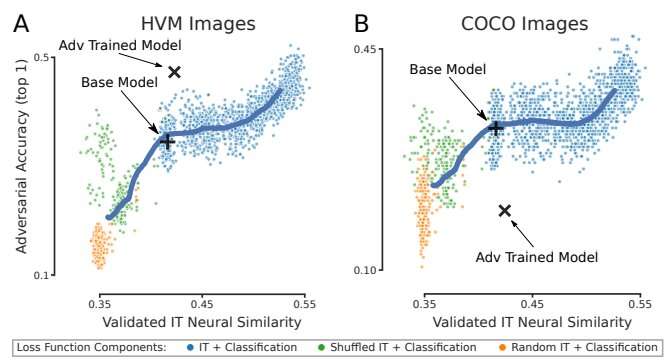
IT neural similarity correlates with improved white box adversarial robustness. A) held out animal and image IT neural similarity is plotted against white box adversarial accuracy (PGD L∞ ϵ = 1/1020) on the HVM image set measured across multiple training time points for all neural loss ratio conditions, random Gaussian IT target matrix conditions, and image shuffled IT target matrix conditions. B) Like in A but for COCO images. In both plots, the black cross represents the average base model position, the black X marks a CORnet-S adversarially trained on HVM images, and the heavy blue line is a sliding X, Y average of all conditions merely to visually highlight trends. Five seeds for each condition are plotted. Credit: Aligning Model and Macaque Inferior Temporal Cortex Representations Improves Model-to-Human Behavioral Alignment and Adversarial Robustness. https://openreview.net/attachment?id=SMYdcXjJh1q&name=pdf From cameras to self-driving cars, many of today’s technologies depend on artificial intelligence to extract meaning from visual information. Today’s AI technology has artificial neural networks at its core, and most of the time we can trust these AI computer vision systems to see things the way we do—but sometimes they falter. According to MIT and IBM research scientists, one way to improve computer vision is to instruct the artificial neural networks that they rely on to deliberately mimic the way the brain’s biological neural network processes visual images.
Researchers led by MIT Professor James DiCarlo, the director of MIT’s Quest for Intelligence and member of the MIT-IBM Watson AI Lab, have made a computer vision model more robust by training it to work like a part of the brain that humans and other primates rely on for object recognition . This May, at the International Conference on Learning Representations, the team reported that when they trained an artificial neural network using neural activity patterns in the brain’s inferior temporal (IT) cortex, the artificial neural network was more robustly able to identify objects in images than a model that lacked that neural training. And the model’s interpretations of images more closely matched what humans saw, even when images included minor distortions that made the task more difficult. Comparing neural circuits
Many of the artificial neural networks used for computer vision already resemble the multilayered brain circuits that process visual information in humans and other primates. Like the brain, they use neuron-like units that work together to process information. As they are trained for a particular task, these layered components collectively and progressively process the visual information to complete the task—determining, for example, that an image depicts a bear or a car or a tree.
DiCarlo and others previously found that when such deep-learning computer vision systems establish efficient ways to solve visual problems, they end up with artificial circuits that work similarly to the neural circuits that process visual information in our own brains. That is, they turn out to be surprisingly good scientific models of the neural mechanisms underlying primate and human vision .
That resemblance is helping neuroscientists deepen their understanding of the brain. By demonstrating ways visual information can be processed to make sense of images, computational models suggest hypotheses about how the brain might accomplish the same task. As developers continue to refine computer vision models, neuroscientists have found new ideas to explore in their own work.
“As vision systems get better at performing in the real world, some of them turn out to be more human-like in their internal processing. That’s useful from an understanding-biology point of view,” says DiCarlo, who is also a professor of brain and cognitive sciences and an investigator at the McGovern Institute for Brain Research. Engineering a more brain-like AI
While their potential is promising, computer vision systems are not yet perfect models of human vision. DiCarlo suspected one way to improve computer vision may be to incorporate specific brain-like features into these models.
To test this idea, he and his collaborators built a computer vision model using neural data previously collected from vision-processing neurons in the monkey IT cortex—a key part of the primate ventral visual pathway involved in the recognition of objects—while the animals viewed various images. More specifically, Joel Dapello, a Harvard University graduate student and former MIT-IBM Watson AI Lab intern; and Kohitij Kar, assistant professor and Canada Research Chair (Visual Neuroscience) at York University and visiting scientist at MIT; in collaboration with David Cox, IBM Research’s vice president for AI models and IBM director of the MIT-IBM Watson AI Lab; and other researchers at IBM Research and MIT asked an artificial neural network to emulate the behavior of these primate vision-processing neurons while the network learned to identify objects in a standard computer vision task.
“In effect, we said to the network, ‘please solve this standard computer vision task, but please also make the function of one of your inside simulated neural layers be as similar as possible to the function of the corresponding biological neural layer,'” DiCarlo explains. “We asked it to do both of those things as best it could.” This forced the artificial neural circuits to find a different way to process visual information than the standard, computer vision approach, he says.
After training the artificial model with biological data, DiCarlo’s team compared its activity to a similarly-sized neural network model trained without neural data, using the standard approach for computer vision. They found that the new, biologically informed model IT layer was—as instructed—a better match for IT neural data. That is, for every image tested, the population of artificial IT neurons in the model responded more similarly to the corresponding population of biological IT neurons.
The researchers also found that the model IT was also a better match to IT neural data collected from another monkey, even though the model had never seen data from that animal, and even when that comparison was evaluated on that monkey’s IT responses to new images. This indicated that the team’s new, “neurally aligned” computer model may be an improved model of the neurobiological function of the primate IT cortex—an interesting finding, given that it was previously […]
Nootropics are being sold as a ‘mind boost’ – but are they a fad?

Nootropics hit the headlines as smart drugs for students to hit their grades. Now this year, they’re being touted as the everyday wellness essential.
Never heard of them? They’re a form of cognitive enhancing supplement offering to enhance brain function and focus. Sounds appealing, right?—if a little scary.
A-listers including Barack Obama and Hillary Clinton reportedly use them. WH uncovered whether they ’re something you should be popping with your vitamin C or well and truly a wellness fad too far. What are nootropics?
“Nootropics are also known as smart drugs. They’re cognitive enhancers, in other words, they make your brain work better, faster and harder,” says Miguel Toribio-Mateas, Clinical Neuroscientist and Registered Nutritionist mBANT.
It all sounds a tad quick fix-y—but don’t confuse your nootropics. There are two types, the synthetic kind and the natural kind.
Contrary to popular opinion, Toribio-Mateas points out that the latter isn’t necessarily made, rather occur naturally in foods, vitamins and minerals. “They’re not always drugs, rather some are in food supplements. Some are herbal or botanical, in other words, they come from a plant.”
And importantly, they are enhancers, not fixers: nootropics should be taken to enhance memory, focus and brain function, not to fix it.
Sources of nootropics you may have heard of include: Caffeine
St. John’s Wort
Ashwagandha
Zinc
Turmeric
Green tea
White tea
Fish oil
Folate
Vitamins B6
Vitamins B12
What are nootropics made of?
If you’re wondering what’s actually in a nootropic, you’re not alone.
The phrase is an umbrella term for hundreds of different types, however, Toribio-Mateas says most are a blend of micronutrients, adaptogens and herbal extracts derived from food-based substances, like vitamin B6, B12 and folate. All work to reduce the amino acid homocysteine, which is known to be an independent marker of cognitive decline, and claim to counter this by enhancing your cognitive performance, ease stress and increase long-term anti-ageing.
But modern medicine this is not. Nootropics have been around for centuries and taken for a variety of ailments. Botanicals like Rhodiola or Ashwagandha to manage stress
Montmorency cherry as a natural source of melatonin to regulate your nocturnal body clock, enhance sleep quality and contribute to waking up feeling more refreshed
Gotu kola to manage anxiety and anger, effectively “softening the edge” of our emotions.
Ginseng as a provider of stamina
Acetyl L-Carnitine helps maintain healthy cognitive function Do nootropics actually work? Although you can’t generalise for all nootropics as there are many hundreds of different strains, there’s actually plenty of studies indicating their effectiveness. Some studies have found piracetam and noopept to be normalise Alzheimer’s memory loss, modafinil to reduce fatigue and semax and phenibut to improve cognitive function.But medication, whether natural or not, should be approached with guidance. Do talk to your GP or regular health professional before guzzling down brain pills with your hot water and lemon. Why the nootropics boom now? Simply put, women are stretching themselves more than ever before and we’re not talking about in the yoga studios. Never-ending work days (thanks Outloook for iPhone) and busy AF lifestyles that are constantly shared on social media an “always on” culture has been created.Disconnecting from the day is rare rather than regular.This is where smart supplements come in.“Nootropic food supplements work gently and often build up over time,” says Toribio-Mateas. “People taking them often report feeling more energised and focused during the day, and enjoying better quality sleep during the night.”However, not all experts back double dropping brain pills at breakfast. “Just because something is natural or claims to improve your health, does not mean it’s safe,” warns Rosie Stunt, dietician at The Rooted Project.“Before turning to supplements as a quick fix, it’s a good idea to assess your lifestyle first. Ask yourself why are you’re so tired or anxious. Are you getting the basics right in your diet? Eating enough fruit and veg? Limiting your alcohol consumption? Getting enough sleep or managing stress levels?”If you fancy giving them a try, remember, nootropic substances are for adults only. Anyone with a diagnosed medical condition or taking any prescribed medication should talk to their doctor, and seek the advice of a knowledgeable nutrition practitioner. Keep reading: we’ve got a guide to metabolism boosters and the symptoms of IBS.
Why people love tea: A look at some herbal tea recipes for mild depression

Advertisement Brighteon Broadcast News, July 3, 2023 – As we celebrate the BIRTH of our nation, its DEATH draws ever closer Advertisement
Tea has a long history and is a part of many traditions from around the world. What makes it all the more special is its health benefits, endless variations and flavors.
Tea is hydrating. Without milk, tea is more than 90 percent water. Drinking four to six mugs of tea a day is as good for keeping you hydrated as a liter of water, reported the Daily Mail , and researchers also found no negative effects from drinking that amount of tea.
Herbal teas and black tea, especially, are great sources of potassium, which plays a central role in making sure your cells can take in the precise amount of water they need – and hence help hydrate your body. (Related: STUDY: Green tea, black tea and matcha tea found to suppress dioxin toxicity .)
In addition, the polyphenols, amino acids and vitamins present in the tea leaves ensure the increase of saliva production – which all contribute to a thirst-quenching feeling.
Tea makes you stronger. Tea contains polyphenols, catechins and biotin which boost and strengthen the immune system. Their antioxidant effects include scavenging reactive oxygen species, inhibiting the formation of free radicals and lipid peroxidation.
These elements protect your cells from free radicals, therefore protecting against blood clots, cancer, or the hardening of the arteries.
Vitamin D found in tea helps build stronger bones, while the amino acids help your body build muscle, as well as fight bad bacteria and viruses.
Tea relaxes you. Tea contains an amino acid that produces a calming effect resulting in a better mood. Also, theanine found in tea reduces anxiety and calms you by increasing the number of inhibitory neurotransmitters.
Researchers have found, for instance, that drinking tea lowers levels of the stress hormone cortisol. And evidence of long-term health benefits is emerging, too – drinking at least 100 milliliters (about half a cup) of green tea a day seems to lower the risk of developing depression and dementia .
For centuries, people across the globe have testified to the relaxing and invigorating qualities of tea. The traditional calming effects of the plant Camellia sinensis have elevated the drink, which is produced from its leaves to a role beyond quenching thirst – people drink tea as an aid for meditation, to help soothe the nerves or SIMPLY TO UNWIND. The magic of a quiet moment: A tip to counter depression
To give your heart and mind time to catch up and process the things that are happening in this world and figure out what role you’re meant to play in it, find the quiet and allow yourself these moments of calm with a cup of tea.
These moments could help you relax better, sleep more deeply, give your days more structure when you’re feeling chaotic, and enhance your overall resiliency.
“The simple act of preparing yourself a cup of tea is, in and of itself, time spent in self-care. Inviting ritual moments into your every day is to call a little bit of calm into the chaos ,” says kitchen herbalist Sass Ayres, plant foods and medicine educator.
Before bed is a great time to sip on a cup of calming herbal tea. Your moment of calm could last five minutes, 20 minutes, an hour, or as long as it takes you to close your eyes and take a single deep breath. Done. The calm that comes with allowing ourselves to feel the freedom in just simply being, even if for a second, is medicine for your whole self. How to make anti-anxiety herbal tea
For equipment, you’ll need: Measuring spoons
Storage containers, such as glass jars
Small saucepan or tea kettle
Tea basket/infuser/strainer
Your favorite mug
Ingredients:
Blend #1 – Un-nerve and nourish tea Chamomile, dried
Lavender, dried
Lemon balm, dried
Oatstraw, dried
Rose hips, dried Blend #2 – Lemon-lavender tea Lavender flowers, dried Lemon peel, dried (or a squeeze of fresh lemon juice) Oatstraw, dried Blend #3 – Field of flowers tea Chamomile flowers, dried Lavender flowers, dried Passion flower, dried Rose petals, dried Blend #4 – Have a good night’s sleep tea Chamomile flowers, dried Peppermint leaves, dried, cut and sifted Rose petals, dried Stinging nettle, dried, cut and sifted Method:Six tablespoons of dried herbal tea make for about six cups (8 fluid ounces) of prepared tea. Make smaller batches first to make sure you like the tea before making it in bulk.You can always tweak your tea by changing the ratios of herbs used or adding in other different calming herbs to suit your taste. > Make the herbal tea blend of your choice. Combine all dried herbs together in a small bowl and mix thoroughly. Boil water. Use a tea kettle or a small saucepan to bring water to a boil. Once boiling, remove from heat. Steep herbs. Using approximately one tablespoon of an anti-anxiety tea blend per 8 fl oz. of water, add your herbs to a tea ball, reusable tea bag, small French press, or other tea strainers. Then add ~8 fl. oz. water just under boiling to your cup. Cover your cup and let steep for 8-10 minutes. The longer you allow the herbs to steep, the stronger your tea will be. Strain & enjoy. After 8-10 minutes, remove the herbs by either straining or removing the tea bag/ball. About the herbs used in these anti-anxiety tea blends These teas are blends of incredibly nourishing and calming plant medicines. Chamomile – is widely regarded as a mild tranquilizer and sleep-inducer . The sedative effects may be due to the flavonoid, apigenin that binds to benzodiazepine receptors in the brain. Studies in preclinical models have shown anticonvulsant and CNS depressant effects respectively. Lavender – helps calm brain function by triggering chemical reactions in the nervous system. Lavender tea boosts the production of dopamine and reduces the stress hormone known as cortisol, according to multiple studies. Lemon balm – essential oils made from lemon […]
Polyphenols in wild blueberries can help lower blood pressure and boost brain function

Advertisement Spokesperson for Vivek Ramaswamy’s presidential campaign joins Brighteon’s Mike Adams for UNCENSORED talk about how to rescue America Advertisement
Wild blueberries ( Vaccinium angustifolium ) are a fixture on the list of the world’s most widely recognized superfoods. Nutritious and delicious, blueberries contain high amounts of vitamins, minerals and dietary fiber, and are one of the richest dietary sources of antioxidant polyphenols . A half-cup serving of ripe wild blueberries (approx. 150 pieces) can provide 200 to 400 milligrams (mg) of polyphenols.
Polyphenols refer to a group of active compounds found naturally in plants, particularly those with vibrant hues. They are extensively studied for their beneficial properties , which include antioxidant, anti-inflammatory, anticancer and anti-diabetic activities.
According to a new study published in the American Journal of Clinical Nutrition (AJCN), the polyphenols in wild blueberries can also boost brain function and help maintain a healthy heart . They recommend consuming 178 grams (about 3/4 cup) of wild blueberries daily for older adults to reduce their risks of cardiovascular disease and cognitive decline. (Related: Study: Regular consumption of blueberries can reverse cognitive decline among the elderly .) Blueberries’ heart benefits linked to improved blood vessel function
Numerous studies have linked the intake of polyphenol-rich blueberries to improvements in vascular function. One notable study involving healthy male volunteers found that consuming wild blueberries daily for one month increased flow-mediated dilation (FMD) , or the widening of coronary arteries due to increased blood flow. This improvement in blood vessel function led to a reduction in ambulatory systolic blood pressure among the participants.
According to the study, polyphenols, specifically anthocyanins, are responsible for the blood pressure-lowering effect of blueberries. Anthocyanins are the pigments found in the skin of blueberries that give them their distinctive blue-purple color. Aside from being natural dyes, anthocyanins are remarkable antioxidants that can effectively neutralize free radicals , thus preventing oxidative stress and inflammation, which are both heavily implicated in the development of many chronic diseases.
In the AJCN study, researchers recruited 61 healthy older adults and randomly assigned them to either the blueberry group or the control group. The former received 26 grams of freeze-dried wild blueberry powder containing 302 mg of anthocyanins, while the latter received a matched placebo that lacked anthocyanins. They then analyzed changes in blood vessel function by looking at FMD, arterial stiffness and blood pressure before and after the 12-week trial.
The researchers reported that, compared with the placebo group, the blueberry group enjoyed a significant increase in FMD and reductions in their 24-hour ambulatory systolic blood pressure. They also noted that total 24-hour urinary polyphenol excretion significantly increased in the blueberry group. These findings suggest that daily intake of blueberry polyphenols can improve blood vessel function in older adults, whose arteries naturally stiffen with age. Arterial stiffening due to aging increases the risk of cardiovascular disease as it limits the ability of the coronary arteries to dilate, causing strain on the heart.
According to studies, anthocyanins are a natural solution to arterial stiffening . They exert their beneficial effects on blood vessels via three mechanisms : They modulate the expression and activity of endothelial nitric oxide synthase, the key enzyme involved in the production of the vasodilating chemical, nitric oxide.
They protect NO from free radicals called reactive oxygen species, which can convert it into a compound that promotes vasoconstriction and hypertension.
They inhibit specific pathways and enzymes involved in the synthesis of vasoconstricting molecules such as angiotensin II, endothelin-I and thromboxanes.
Blueberry polyphenols can improve brain function
Aside from lowering blood pressure, consuming wild blueberries has also been linked to enhanced brain performance. The results of randomized clinical trials suggest that blueberry consumption or supplementation can improve various measures of cognitive performance , particularly short- and long-term memory and spatial memory. There is also evidence that adding blueberries to your diet can positively influence your mood.
Just like with blood vessels, improvements in brain function after short- or long-term intake of blueberries are credited to their anthocyanin content. According to a study published in the Journal of Agricultural and Food Chemistry , the anthocyanins in blueberries can mitigate neurodegeneration by increasing neuronal signaling in brain centers , which explains why blueberry consumption has been found to enhance memory function in clinical trials.
After 12 weeks of drinking wild blueberry juice daily, the study reported that older adults with early memory changes experienced improvements in paired associate learning and word list recall. The researchers also noted that drinking blueberry juice reduced depression symptoms among the participants and lowered their blood sugar levels.
In the AJCN study, the researchers also investigated whether the cognitive benefits of consuming blueberries are linked to increases in cerebral blood flow and vascular blood flow as well as changes in gut microbiota. While they observed an increase in FMD, they found no changes in cerebral blood flow or the gut microbial composition of the participants who were supplemented with freeze-dried wild blueberry powder for 12 weeks.
On the other hand, they noticed significant improvements in cognitive function , specifically enhanced immediate recall on the auditory verbal learning task and better accuracy on the task-switch task, which measures how fast a person can shift his attention from one task to the other. Based on these results, the researchers concluded that daily consumption of blueberries can not only protect against cardiovascular disease but also improve episodic memory processes and executive functioning in older adults who are at risk of cognitive decline. ( Related: Blueberries: Superfood that can help you improve brain health .) Other health benefits of eating blueberries
Antioxidant-rich blueberries are known to provide a wide range of health benefits . According to studies, a well-balanced diet that includes fresh, organic blueberries can help you: Maintain strong and healthy bones , thanks to the bone-supporting minerals (i.e., calcium, iron, magnesium, manganese, phosphorus and zinc) and vitamin K in blueberries
Maintain healthy, younger-looking skin , thanks to blueberries’ high vitamin C content, which helps protect against skin damage and increases collagen synthesis
Maintain healthy blood sugar levels , […]
Len Rome’s Local Health: Menopause and memory loss

(WYTV)- Here’s what doctors are telling us about menopause today: there’s a connection between it and some memory loss, or call it a kind of brain fog.
We know that menopause can increase the risk for heart disease and osteoporosis…and now memory.
Women often admit they’re worried during menopause when their memories seem to slip. And studies have confirmed this brain fog.
“Multiple studies have shown cognitive complaints by women that are going through the menopause transition both subjectively like, oh gosh, I keep forgetting where my keys are.
But also, objectively when they do cognitive tests, they see changes in executive function.”
The good news, brain fog appears to be temporary, tests for it after menopause transition do show improvement. Hot flashes, night sweats, weight gain, and now brain fog.
Check with your doctor to find out what treatment is right for you.
Copyright 2023 Nexstar Media Inc. All rights reserved. This material may not be published, broadcast, rewritten, or redistributed.

The Power Within by Corey Daniels book available for only $2.99
Mind has both conscious and subconscious halves. These are likened to a driver and the truck he drives. The driver plans the destination and observes road conditions, while the truck provides motive power. Your subconscious mind is like the truck and it only goes in the direction in which is a steered. This can be the road or off a cliff. Likewise, the consciousness paints a picture of what the world is and what your goals are and the subconscious acts on them through emotion, physical response and energy, whether these are correct, rational images or false negative ones. The subconscious is also like an emotional reservoir which your body and mind draw responses from to external stimuli.
The Power Within lays out a method of programming your subconscious and tapping into the Holy Spirit, God voice or what the Greeks called the daimon (godman).