Nature Knows and Psionic Success
God provides
STUDY: Short-term face mask use causes carbon dioxide poisoning – cognitive impairment, testicular damage, stillbirth and impaired memory

( Natural News ) Studies continue to show that face masks provided no protection against covid-19. Now, a new peer-reviewed study out of Germany finds that short-term face mask use causes carbon dioxide poisoning, leading to testicular dysfunction in young men, increased risk of stillbirth for pregnant women, cognitive decline in children, as well as impaired memory, anxiety, and other serious health problems. Masks force a person to inhale unsafe levels of CO2, synthetic microfibers, carcinogenic compounds, volatile organic compounds, and microorganisms that have adapted to the moist environment inside the mask. The researchers warn that face masks suffocate people in their own exhaled waste, increasing CO2 levels in their blood while driving up blood pressure and inflammatory markers.
The study, Possible toxicity of chronic carbon dioxide exposure associated with face mask use, particularly in pregnant women, children, and adolescents was published in the peer reviewed journal, Helion . Face masks spike CO2 blood levels, destroying childhood brain development, depleting men’s sperm
Fresh air typically contains just .04% CO2. When a person puts on a mask, they are exposed to low level carbon dioxide poisoning in the range of 1.4–3.2%. In the study, CO2 concentrations as low as .3% were associated with significant brain damage, impaired memory, and increased anxiety in children. The study found that just five minutes of mask wearing can expose an individual to dangerous CO2 levels – laying the groundwork for serious health issues and developmental disorders in children.
In one study , 0.3% CO2 exposure on adolescent brain neurons “can cause destruction in the gyrus dentatus and the prefrontal cortex with decreased IGF-1 levels resulting in less activity, increased anxiety and impaired learning and memory.”
The concentration of CO2 in the blood has an important influence on intra- and extracellular pH. When CO2 passes quickly through the cell membranes, it goes on to form carbonic acid with H2O, causing the release of H + ions, leading to acidosis and the die-off of neurons.
When male mice are exposed to 2.5% CO2 for four hours, their testicular cells and sperm are destroyed. For humans, this exposure is equal to .5% CO2 — a common exposure during mask mandates. In the study, four hours of low-level CO2 exposure causes spermatid and Sertoli cells in testes to self-destruct, causing streaking & vacuolization of the tubular components and consequentially no maturation of the spermatids. CO2 poisoning of pregnant women causes birth defects, higher risk of stillbirth
Carbon dioxide exposure can cause oxidative stress and the formation of lipid hydroperoxides that cause DNA fragmentation and subsequent mitochondrial damage and cell death. Pregnant rats exposed to 3% CO2 were more likely to suffer stillbirths and have offspring with birth defects. This is equal to 0.8 percent CO2 exposure for humans – a common exposure for pregnant women who worked under mask mandates.
Before widespread mask use, the stillbirth rate in humans was 7 per 1000 births. During the masking pandemonium, stillbirths increased to 21 per 1,000 births. A prominent UK hospital reported a fourfold increase in stillbirths during the covid-19 scandal, and carbon dioxide poisoning was a major contributing factor. The damage was also observed in Australia , where people were forced to wear masks for years. These increase in stillbirths was not observed in Sweden , where there were no mask mandates.
“Circumstantial evidence exists that popular mask use may be related to current observations of a significant rise of 28 percent to 33 percent in stillbirths worldwide,” the German researchers concluded.
Forceful and coercive face mask policies continue to violate the sovereign rights and health of the individual, weakening their immune system and setting up their oxygen-deprived cells for oxidative damage, inflammation, and severe disease. Pregnant women and their fetuses were directly harmed by policies of forced CO2 poisoning. Forced masking of children caused negative psychological effects and additional physiological damage to their brains, their immune system, and their development.
Sources include:
BMCPublicHealth.BioMedCentral.com
Cell.com [PDF]
ScienceDirect.com
TandFOnline.com
Rep.Bioscientifica.com
Summit.news
MedRXIV.org
ACPJournals.org
NaturalNews.com
Music therapy may help children with brain injuries: Research

Representative Image . Image Credit: ANI New pilot research has found that a music therapy strategy may help youngsters with severe acquired brain injuries (ABI) meet their walking recovery goals. The first of its kind research project suggests rhythmic auditory stimulation could augment the physiotherapy ordinarily offered to children and young people who sustain a severe acquired brain injury – of whom there are believed to be around 350 each year in the UK.
The practice aims to improve walking speed and quality of movement by using rhythms to provide cues for patients’ stepping rate. In establishing neurological connections between the auditory prompts and physical movements, the technique has been shown to facilitate smoother and more coordinated walking patterns. While the potential of rhythmic auditory stimulation has never before been explored in the context of treating children with ABIs, the approach has been found to improve walking speed among people who have suffered a stroke, those living with Parkinson’s disease and, more recently, children with cerebral palsy.
This is a modal window.
No compatible source was found for this media. All participants in the pilot study, published in the International Journal of Therapy and Rehabilitation and involving a music therapy expert from Anglia Ruskin University (ARU), showed improvement as a result of rhythmic auditory stimulation being added to their rehabilitation programme, with the most marked advances evident in walking quality. In light of these findings, the study’s authors believe adding the technique to other interventions could help improve the outlook for children with ABIs.
Dr Jonathan Pool, Senior Research Fellow at Anglia Ruskin University’s Cambridge Institute for Music Therapy Research and an author of the study, said: “This is the first study to look at rhythmic auditory stimulation for children and young people with acquired brain injury. “As a pilot study, it provides initial evidence of the effect of music on gait rehabilitation for this population and has revealed insights into some of the issues for researchers in this area.
“While showing variation across participants in the benefits of rhythmic auditory stimulation, the study findings are encouraging and indicate that the detailed assessment of quality of movement should be considered alongside other tests when measuring functional gains in gait rehabilitation.” (ANI)
(This story has not been edited by Devdiscourse staff and is auto-generated from a syndicated feed.)
Biological Brains Outpace AI in Learning, Thanks to Structured Exploration
Summary: When it comes to rapid learning, the biological brain seems to have an edge over current AI technology.
Scientists worked with different models of reinforcement learning to understand the algorithms that the brain uses to learn. They found that the directed exploration used by animals makes learning more efficient and requires less experience, unlike artificial agents that explore randomly.
Key Facts:
> The study suggests that animals’ directed exploration makes learning more efficient, which could help build better AI agents that can learn faster and require less experience.
AI agents need a lot of experience to learn something and explore the environment thousands of times, whereas a real animal can learn an environment in less than ten minutes.
The research findings emphasize the need for more efficient learning algorithms that can explain the behavior of animals in exploring and learning their spatial environment.
Source: Sainsbury Wellcome Center
Neuroscientists have uncovered how exploratory actions enable animals to learn their spatial environment more efficiently. Their findings could help build better AI agents that can learn faster and require less experience.
Researchers at the Sainsbury Wellcome Centre and Gatsby Computational Neuroscience Unit at UCL found the instinctual exploratory runs that animals carry out are not random. These purposeful actions allow mice to learn a map of the world efficiently.
The study, published today in Neuron , describes how neuroscientists tested their hypothesis that the specific exploratory actions that animals undertake, such as darting quickly towards objects, are important in helping them learn how to navigate their environment.
“There are a lot of theories in psychology about how performing certain actions facilitates learning. In this study, we tested whether simply observing obstacles in an environment was enough to learn about them, or if purposeful, sensory-guided actions help animals build a cognitive map of the world,” said Professor Tiago Branco, Group Leader at the Sainsbury Wellcome Centre and corresponding author on the paper.
In previous work, scientists at SWC observed a correlation between how well animals learn to go around an obstacle and the number of times they had run to the object. In this study, Philip Shamash, SWC PhD student and first author of the paper, carried out experiments to test the impact of preventing animals from performing exploratory runs.
By expressing a light-activated protein called channelrhodopsin in one part of the motor cortex, Philip was able to use optogenetic tools to prevent animals from initiating exploratory runs towards obstacles.
The team found that even though mice had spent a lot of time observing and sniffing obstacles, if they were prevented in running towards them, they did not learn. This shows that the instinctive exploratory actions themselves are helping the animals learn a map of their environment.
To explore the algorithms that the brain might be using to learn, the team worked with Sebastian Lee, a PhD student in Andrew Saxe’s lab at SWC, to run different models of reinforcement learning that people have developed for artificial agents, and observe which one most closely reproduces the mouse behaviour. There are two main classes of reinforcement learning models: model-free and model-based. Credit: Neuroscience News There are two main classes of reinforcement learning models: model-free and model-based. The team found that under some conditions mice act in a model-free way but under other conditions, they seem to have a model of the world. And so the researchers implemented an agent that can arbitrate between model-free and model-based. This is not necessarily how the mouse brain works, but it helped them to understand what is required in a learning algorithm to explain the behaviour.
“One of the problems with artificial intelligence is that agents need a lot of experience in order to learn something. They have to explore the environment thousands of times, whereas a real animal can learn an environment in less than ten minutes.
“We think this is in part because, unlike artificial agents, animals’ exploration is not random and instead focuses on salient objects. This kind of directed exploration makes the learning more efficient and so they need less experience to learn,” explains Professor Branco.
The next steps for the researchers are to explore the link between the execution of exploratory actions and the representation of subgoals. The team are now carrying out recordings in the brain to discover which areas are involved in representing subgoals and how the exploratory actions lead to the formation of the representations.
Funding: This research was funded by a Wellcome Senior Research Fellowship (214352/Z/18/Z) and by the Sainsbury Wellcome Centre Core Grant from the Gatsby Charitable Foundation and Wellcome (090843/F/09/Z), the Sainsbury Wellcome Centre PhD Programme and a Sir Henry Dale Fellowship from the Wellcome Trust and Royal Society (216386/Z/19/Z). About this AI and neuroscience research news
Author: April Cashin-Garbutt
Source: Sainsbury Wellcome Center
Contact: April Cashin-Garbutt – Sainsbury Wellcome Center
Image: The image is credited to Neuroscience News
Original Research: Open access.
“ Mice identify subgoal locations through an action-driven mapping process ” by Sebastian Lee et al. Neuron
Abstract
Mice identify subgoal locations through an action-driven mapping process Highlights
Interruption of obstacle-directed runs during exploration prevents subgoal learning Subgoal selection during escape depends on the mouse’s position in the environment A dual-system reinforcement learning agent replicates the mouse behavior Summary Mammals form mental maps of the environments by exploring their surroundings. Here, we investigate which elements of exploration are important for this process. We studied mouse escape behavior, in which mice are known to memorize subgoal locations—obstacle edges—to execute efficient escape routes to shelter.To test the role of exploratory actions, we developed closed-loop neural-stimulation protocols for interrupting various actions while mice explored. We found that blocking running movements directed at obstacle edges prevented subgoal learning; however, blocking several control movements had no effect.Reinforcement learning simulations and analysis of spatial data show that artificial agents can match these results if they have a region-level spatial representation and explore with object-directed movements. We conclude that mice employ an action-driven process for integrating subgoals into a hierarchical cognitive map.These […]

This Is Your Brain on Books: Science Reveals Secrets of Reading

MONDAY, April 24, 2023 (HealthDay News) — Reading is fundamental, but it’s also a complex skill. Now, a new study sheds more light on how the brain makes sense of the written word.
Researchers found that two key brain “networks” work in tandem to help people read sentences — so folks not only grasp the meanings of individual words but also process the bigger picture of what’s being said.
Because reading is such an essential daily activity, it’s easy to take it for granted, said study leader Oscar Woolnough , a research fellow with McGovern Medical School at UTHealth Houston.
“That is, until you lose that ability,” he said.
Woolnough pointed to the example of aphasia, which impairs people’s ability to use language — including their speech and ability to write or read. It stems from damage to the brain, often from a stroke or a head injury. People are also reading…
Friends identify man killed in fire as revered St. Louis blues musician Tom Hall
BenFred: Cardinals were right to not trade for Diamondbacks’ Madison Bumgarner
Can ‘fiery’ Jack Flaherty’s controlled burn of emotions be what ignites Cardinals?
Wash U. says ex-employee’s claims about transgender center in St. Louis are unfounded
If researchers can better understand how the healthy brain allows people to read, Woolnough said, that could improve understanding of aphasia and other types of reading impairment.
For the latest study, the researchers recruited patients with epilepsy who’d had electrodes implanted in their brains to try to identify the source of their seizures.
That allowed Woolnough’s team to record the participants’ brain activity as they read — precisely charting the timing of events in a way not possible with noninvasive imaging of the brain.
Researchers had the 36 participants silently read various sentences and word lists — some composed of real words and some composed of nonsense “Jabberwocky” words (based on Lewis Carroll’s ” Jabberwocky ” poem).
It turned out that when people were reading real sentences, two distinct networks in the brain’s frontotemporal cortex jumped into action. In the first one, activity progressively increased as readers absorbed sentences — a ramping up not seen when people read a word list.
That, Woolnough explained, suggests the network is adding up the combined meaning of the individual words in a sentence, and building the bigger picture of what’s being said.
The second network the researchers identified worked differently: It was more active when people were reading word lists, versus sentences. But that’s not because it was lazy during sentence reading.
As Woolnough explained it, the second network seems to become more efficient when people are reading sentences — because the context of the sentence makes it easier to process the individual words.
“Your brain can start to predict what’s coming next,” he said.
The findings — published recently in the Proceedings of the National Academy of Sciences — may not have any immediate implications for addressing reading disorders.
But experts said the study highlights the complexity of a task that is vital to everyday life.
Reading ability cannot be pinpointed to any one hub in the brain, said Monica McQuaid , director of the adult literacy program at Montefiore Medical Center’s Fisher Landau Center for the Treatment of Learning Disabilities in Bronx, N.Y.
Instead, it involves an orchestration of activity from various areas of the brain.
Dyslexia, for instance, sometimes gets misunderstood as a disorder where people see words “backwards,” McQuaid said. But the issue is not a visual one, she explained. It’s one of language processing.
So addressing dyslexia takes a “multi-sensory” approach, McQuaid explained. Instead of, say, just showing a child the word “cat,” a therapist can also use a picture of a cat, the recorded sound of a cat or integrate the movement of a cat — to “build meaning.”
When it comes to aphasia, people commonly think it’s a speech impairment, said Sarah Wallace , a professor of communication science and disorders at the University of Pittsburgh.
In reality, she said, aphasia impacts all language processing — speaking, writing and reading.Like McQuaid, Wallace pointed to the array of brain areas involved in reading, and the need for “multi-faceted” approaches to managing impairments.There are therapies to help people with aphasia improve their reading abilities, generally involving reading aloud. At the same time, Wallace said, it’s also important to make the task easier: Technology is one way to help, with text-to-speech devices that present written text and computerized speech at the same time, for example.Technology, though, has also made everyone more reliant on reading, Wallace pointed out. Emails and text messages have replaced the old-fashioned phone call.”Reading is such a huge part of our everyday lives,” Wallace said. “It’s part of how we make new relationships, and sustain relationships.”So it’s “critically important,” she said, to better understand such a fundamental human ability. More information The American Speech-Language Hearing Association has more on aphasia .SOURCES: Oscar Woolnough, PhD, postdoctoral research fellow, Department of Neurosurgery, McGovern Medical School at UTHealth Houston; Sarah Wallace, PhD, CCC-SLP, professor, communication science and disorders, School of Health & Rehabilitation Sciences, University of Pittsburgh; Monica McQuaid, PhD, director, Adult Literacy Program, Fisher Landau Center for the Treatment of Learning Disabilities, Children’s Evaluation and Rehabilitation Center at Montefiore, Bronx, N.Y.; Proceedings of the National Academy of Sciences , April 17, 2023, online 0 Comments Tags Dcc Wire Tncen Health-and-wellness Healthday Consumer News Originally published on consumer.healthday.com , part of the BLOX Digital Content Exchange .
Scientists Identify Brain Aging “Sweet Spot”
Indigenous communities in lowland Bolivia, such as the Tsimané and Mosetén, have some of the lowest rates of heart and brain disease due to optimal levels of food consumption and exercise. New research indicates that these communities’ lifestyles, which balance daily exertion and food abundance, contribute to healthy brain aging and reduced risk of disease. Indigenous communities residing in the tropical forests of lowland Bolivia have reported some of the lowest rates of heart disease and brain disease in recorded scientific history. Now, research conducted by USC on the Tsimané and Mosetén communities indicates that a balanced combination of food consumption and physical activity can maximize healthy brain aging and decrease the likelihood of disease.
The study was recently published in the journal Proceedings of the National Academy of Sciences .
The advent of industrialization has brought about numerous benefits, including increased availability of food, reduced physical toil, and improved healthcare access. However, our current way of life has also resulted in a lack of exercise and overconsumption of food, leading to the rise of obesity. Unfortunately, this sedentary lifestyle and obesity are linked to smaller brain volumes and quicker cognitive decline.
To better understand the tipping point where abundance and ease begin to undermine health, the researchers enrolled 1,165 Tsimané and Mosetén adults, aged 40-94 years, and provided transportation for participants from their remote villages to the closest hospital with CT scanning equipment.
The Tsimané have some of the lowest rates of heart and brain disease in the world. Credit: Tsimane Health and Life History Project Team
The team used CT scans to measure brain volume by age. They also measured participants’ body mass index, blood pressure, total cholesterol, and other markers of energy and overall health.
Researchers found that the Tsimané and Mosetén experience less brain atrophy and improved cardiovascular health compared to industrialized populations in the U.S. and Europe. Rates of age-related brain atrophy, or brain shrinking, are correlated with risks of degenerative diseases like dementia and Alzheimer’s.
“The lives of our pre-industrial ancestors were punctured by limited food availability,” said Andrei Irimia, an assistant professor of gerontology, biomedical engineering, quantitative/computational biology, and neuroscience at the USC Leonard Davis School of Gerontology and co-corresponding author of the study. “Humans historically spent a lot of time exercising out of necessity to find food, and their brain aging profiles reflected this lifestyle.” The Mosetén: A bridge between pre- and post-industrialized societies
The findings also illustrated key differences between the two Indigenous societies. The Mosetén are a “sister” population to the Tsimané in that they share similar languages, ancestral history, and a subsistence lifestyle. However, the Mosetén have more exposure to modern technology, medicine, infrastructure, and education.
“The Mosetén serve as an important intermediary population that allows us to compare a wide spectrum of lifestyle and health care factors. This is more advantageous than a straight comparison between the Tsimané and the industrialized world,” Irimia said.
Irimia said that, along this continuum, the Mosetén showed better health than modern populations in Europe and North America — but not as good as that of the Tsimané.
Among the Tsimané, surprisingly, BMI and somewhat higher levels of “bad cholesterol” were associated with bigger brain volumes for age. This, however, may be due to individuals being more muscular, on average, than individuals in industrialized countries who have comparable BMIs.
Still, both the Tsimané and Mosetén come closer to the “sweet spot,” or balance between daily exertion and food abundance, that the authors think may be key to healthy brain aging. The future of preventative medicine relies on an understanding of humans’ evolutionary past
The study’s authors explained that people living in societies with abundant food and little requirement for physical activity face a conflict between what they consciously know is best for their health and the cravings, or drives, that come from our evolutionary past.
“During our evolutionary past, more food and less calories spent in getting it resulted in improved health, well-being and ultimately higher reproductive success or Darwinian fitness,” notes Hillard Kaplan, a professor of health economics and anthropology at Chapman University who has studied the Tsimané for nearly two decades. “This evolutionary history selected for psychological and physiological traits that made us desirous of extra food and less physical work, and with industrialization, those traits lead us to overshoot the mark.”
According to Irimia, the best place to be in terms of brain health and risk for disease is the “sweet spot” where the brain is being provided with neither too little nor too much food and nutrients, and where you have a vigorous amount of exercise.
“This ideal set of conditions for disease prevention prompts us to consider whether our industrialized lifestyles increase our risk of disease,” he said.
Reference: “Brain volume, energy balance, and cardiovascular health in two nonindustrial South American populations” by Hillard Kaplan, Paul L. Hooper, Margaret Gatz, Wendy J. Mack, E. Meng Law, Helena C. Chui, M. Linda Sutherland, James D. Sutherland, Christopher J. Rowan, L. Samuel Wann, Adel H. Allam, Randall C. Thompson, David E. Michalik, Guido Lombardi, Michael I. Miyamoto, Daniel Eid Rodriguez, Juan Copajira Adrian, Raul Quispe Gutierrez, Bret A. Beheim, Daniel K. Cummings, Edmond Seabright, Sarah Alami, Angela R. Garcia, Kenneth Buetow, Gregory S. Thomas, Caleb E. Finch, Jonathan Stieglitz, Benjamin C. Trumble, Michael D. Gurven and Andrei Irimia, 20 March 2023, Proceedings of the National Academy of Sciences .
DOI: 10.1073/pnas.2205448120
The study was funded by the National Institute on Aging, the National Science Foundation, and the French National Research Agency – Investissements d’Avenir.

Exercise boosts brain health with chemical signals, study shows
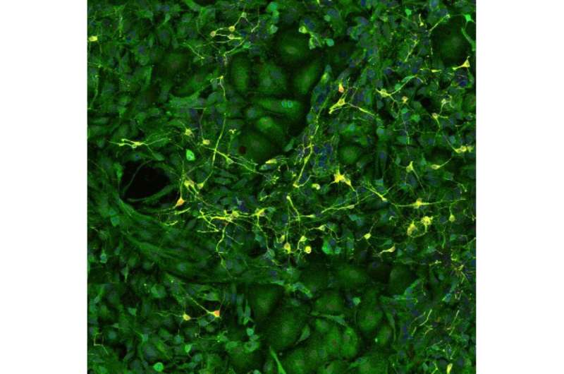
Hippocampal neurons (yellow) surrounded by astrocytes (green) in a cell culture from the study. Image provided by the authors. Credit: Taher Saif, Justin Rhodes, and Ki Yun Lee Physical activity is frequently cited as a means of improving physical and mental health. Researchers at the Beckman Institute for Advanced Science and Technology have shown that it may also improve brain health more directly. They studied how the chemical signals released by exercising muscles promote neuronal development in the brain.
Their work appears in the journal Neuroscience .
When muscles contract during exercise , like the biceps working to lift a heavy weight, they release a variety of compounds into the bloodstream. These compounds can travel to different parts of the body, including the brain. The researchers were particularly interested in how exercise could benefit a particular part of the brain called the hippocampus.
“The hippocampus is a crucial area for learning and memory, and therefore cognitive health,” said Ki Yun Lee, a Ph.D. student in mechanical science and engineering at the University of Illinois Urbana-Champaign and the study’s lead author. Understanding how exercise benefits the hippocampus could therefore lead to exercise-based treatments for a variety of conditions including Alzheimer’s disease.
To isolate the chemicals released by contracting muscles and test them on hippocampal neurons , the researchers collected small muscle cell samples from mice and grew them in cell culture dishes in the lab. When the muscle cells matured, they began to contract on their own, releasing their chemical signals into the cell culture.
The research team added the culture, which now contained the chemical signals from the mature muscle cells, to another culture containing hippocampal neurons and other support cells known as astrocytes . Using several measures, including immunofluorescent and calcium imaging to track cell growth and multi-electrode arrays to record neuronal electrical activity, they examined how exposure to these chemical signals affected the hippocampal cells.
The results were striking. Exposure to the chemical signals from contracting muscle cells caused hippocampal neurons to generate larger and more frequent electrical signals —a sign of robust growth and health. Within a few days, the neurons started firing these electrical signals more synchronously, suggesting that the neurons were forming a more mature network together and mimicking the organization of neurons in the brain.
However, the researchers still had questions about how these chemical signals led to growth and development of hippocampal neurons. To uncover more of the pathway linking exercise to better brain health, they next focused on the role of astrocytes in mediating this relationship.
“Astrocytes are the first responders in the brain before the compounds from muscles reach the neurons,” Lee said. Perhaps, then, they played a role in helping neurons respond to these signals.
The researchers found that removing astrocytes from the cell cultures caused the neurons to fire even more electrical signals, suggesting that without the astrocytes, the neurons continued to grow—perhaps to a point where they might become unmanageable.
“Astrocytes play a critical role in mediating the effects of exercise,” Lee said. “By regulating neuronal activity and preventing hyperexcitability of neurons, astrocytes contribute to the balance necessary for optimal brain function.”
Understanding the chemical pathway between muscle contraction and the growth and regulation of hippocampal neurons is just the first step in understanding how exercise helps improve brain health.
“Ultimately, our research may contribute to the development of more effective exercise regimens for cognitive disorders such as Alzheimer’s disease,” Lee said.
In addition to Lee, the team also included Beckman faculty members Justin Rhodes, a professor of psychology; and Taher Saif, a professor of mechanical science and engineering.
Provided by Beckman Institute for Advanced Science and Technology
8 Science-backed health benefits of eating walnuts
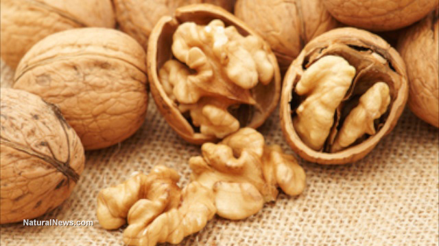
( Natural News ) Walnuts, with their hard shells, are the healthiest of all nuts and should be eaten as part of a healthy diet. According to researchers from Pennsylvania, walnuts contain the highest concentration of antioxidants among known nuts.
Dr. Joe Vinson from the University of Scranton discovered that a handful of walnuts contain twice as many antioxidants as a handful of any other commonly eaten nut. This makes walnuts excellent sources of beneficial, naturally occurring chemicals that can fight oxidative stress.
Here are eight more reasons why you should eat walnuts every day. Walnuts support liver detoxification
Loaded with omega-3 fatty acids, the master antioxidant glutathione and the amino acid arginine, walnuts can cleanse your liver naturally by detoxifying ammonia. Because they help increase blood circulation, walnuts speed up the delivery of toxins to the liver, the organ that facilitates their elimination from the body.
According to a study published in the Biomedical and Pharmacology Journal , the arginine in walnuts has immune-supporting effects , especially under catabolic conditions. Arginine also helps in the treatment of liver disorders, such as liver cirrhosis and fatty liver disease. Walnuts support heart health
Walnuts are rich in nutrients that are great for your heart, such as omega-3 fatty acids, vitamin E, folate and magnesium.
The healthy omega-3 fats , more specifically alpha-linolenic acid, present in walnuts can help raise your good cholesterol levels, lower bad cholesterol and prevent the development of erratic heart rhythms. Eating walnuts regularly has been proven to help reduce inflammation in the arteries, which can prevent heart disease.
In a study published in the journal Nutrition, Metabolism & Cardiovascular Disease , researchers found that walnut eaters have a better heart disease risk profile , including a smaller waist circumference and lower body mass index, blood pressure and blood triglyceride levels, than non-walnut eaters and those who eat other nuts. Walnuts support brain health
Walnuts are known as “brain food,” but it is not only due to their brain-like shape. Walnuts are rich in healthy fatty acids, iodine and selenium that can improve brain function.
Studies have shown that walnuts can also reduce inflammation in the brain , which is linked to the development of neurodegenerative diseases like Alzheimer’s.
According to a study published in the journal Nutrients , the addition of walnuts in one’s diet helps reduce the risk of mild cognitive impairment and neurodegenerative diseases. Walnuts can keep your gut healthy
Studies suggest that if your gut is rich in health-promoting microbes, you’re more likely to have a healthy gut, a strong immune system and good overall health. Eating walnuts may be one way to support a healthy gut microflora.
A randomized controlled trial published in the journal Nutrients found that consuming 1.5 ounces (43 grams) of walnuts every day helps increase beneficial gut bacteria that butyrate, a short-chain fatty acid that nourishes your gut and promotes good gut health. Walnuts fight cancer
Test-tube, animal and human observational studies, including one published in the journal Gynecologic and Obstetric Investigation , show that walnuts are great at fighting cancer due to their antioxidant properties , which give them the ability to protect the body against breast, colon and prostate cancer.
A study published in the journal Critical Reviews in Food Science and Nutrition reported that walnuts are rich in ellagitannins, which certain gut microbes can convert to compounds called urolithins. The anti-inflammatory properties of urolithins can help lower your risk of developing colorectal cancer. Walnuts can help you lose weight
Research has found that eating a handful of walnuts every day can help stimulate fat loss and promote a healthy weight. Walnuts are known for their ability to suppress appetite , thanks to the presence of omega-3 fatty acids, plant sterols, protein and fiber that help curb hunger. (Related: Walnuts reduce hunger and cravings by changing your brain .)
A study published in the journal Diabetes, Obesity and Metabolism also found that walnut consumption increases the activation of the insula , a brain region involved in appetite and impulse control, to help curb appetite when you see “highly desirable” food. Walnuts support blood sugar control
Walnuts are diabetes-friendly nuts , being low in carbohydrates that could elevate glucose levels. On the other hand, walnuts are high in fiber, protein and healthy fats that could help prevent blood sugar spikes, says dietitian-nutritionist Cheryl Mussato, founder of Eat Well to Be Well in Osage City, Kansas. Walnuts strengthen your bones
Walnuts are a great source of copper, which can neutralize superoxide radicals produced during bone resorption. Walnuts are also rich in magnesium, which improves bone mineral density, and manganese, a co-factor for several enzymes involved in bone formation. The best way to eat walnuts
Walnuts can be eaten directly from their shell. You can soak them overnight and eat them first thing in the morning. Soaking eases digestion, lowers the level of phytic acid in walnuts and promotes metabolism and nutrient absorption.
As nutritious as walnuts are, you don’t need to eat a lot to experience their benefits. Moderate consumption of walnuts has been shown to be the best way to enjoy all the wholesome goodness this superfood has to offer. Snack on seven to 10 walnuts per day or add them to your favorite recipes to stay healthy.
Watch the following video to learn how walnuts can keep your gut healthy . Keep your gut healthy with walnuts
This is a modal window.
No compatible source was found for this media.
This video is from the Natural News channel on Brighteon.com . More related stories:
Eating walnuts found to protect the colon from cancerous tumors . Walnuts are a delicious way to take care of your heart . Help manage stress, blood pressure with walnuts . Sources include: BBC.com BiomedPharmaJournal.org NMCD-jounral.com NCBI.NLM.NIH.gov PubMed.NCBI.NLM.NIH.gov 1 PubMed.NCBI.NLM.NIH.gov 2 PubMed.NCBI.NLM.NIH.gov 3 NDTV.com Wiley.com EverydayHealth.com HealthierSteps.com Brighteon.com

On nutrition: The gut-brain connection
Credit: Pixabay/CC0 Public Domain If you ever thought your digestive system’s only role was to break down food, think again.
According to microbiome researcher Christopher Lowry, “what happens in the gut affects other parts of the body, including the brain.” And much of this activity is related to trillions of good and bad bacteria —collectively called our “microbiome”—that inhabit our gastrointestinal tract.
Lowry, who is currently conducting research at the Department of Integrative Physiology and Center for Neuroscience at the University of Colorado, Boulder on the relationship between stress-related disorders and the bacteria that live in our guts, was a featured speaker at a daylong workshop hosted by the Institute for Brain Potential in Cheyenne, Wyoming, titled “Understanding the Gut Brain.”
“Americans are microbially deprived,” said Lowry. “Gut microbes are critical for a healthy gut and a healthy brain,” and a diversity of good bugs appears to be the most beneficial.
That’s because each type of microbe has a distinct effect on the body. Some produce natural antibiotics to fight infections, for instance. Others enhance our body’s ability to extract energy from food. Still others produce chemicals that can actually improve our moods and calm stress. When the balance of these good bugs gets out of whack, we open ourselves to not-so-good conditions.
So how do we encourage the growth of beneficial bacteria in our guts? Spend more time in nature, Lowry suggested. That’s where we encounter a wide variety of healthful microbes.
And, consume a variety of foods that feed good bacteria.
Top of the list? Plants. Health-promoting microbes thrive on fibers found in foods that sprout from the ground.
“Each plant has its own microbiome,” Lowry explained. And according to results from the American Gut Project, the largest published study to date of the human microbiome, people who ate at least 30 different types of plant foods a week had more diverse bacteria in their guts than those who ate 10 or less.
My husband and I are not vegetarians, but we do consume our fair share of fruit, vegetables, beans and grains. How hard would it be to eat 30 varieties of these foods every week?
We took the challenge. For a week, I recorded every different type of plant food we ate, no matter how much or how little. For example, our salad one night had cabbage, kale, carrots, raisins and sunflower seeds. That’s five!
The next day we had blueberries and bananas in our oatmeal, three more. Celery, onion, beans, corn, tomatoes and garlic in my stew the next day added another six to our list.
It was kind of fun … and not impossible. I found myself searching for olives to toss into egg salad. And each type of nut I ate for a snack contributed to a wider diversity of my microbiome.
The point, according to this and other researchers, is that our bodies thrive on variety. And when it comes to healthful food choices, that may be a critical key to health.
2023 MediaNews Group, Inc.
Distributed by Tribune Content Agency, LLC.
Scientists Enhance New Neurons to Restore Memory and Elevate Mood in Alzheimer’s Disease
Summary: Stimulating the Supramammilary nucleus in the hypothalamus enhanced adult-born neurons in the hippocampus of mouse models of Alzheimer’s disease. These modified adult-born neurons restored both cognitive impairments and mood-related disorders associated with Alzheimer’s in the mice.
Source: UNC
In adult human brains, the hippocampus generates new neurons (adult-born neurons, or ABNs) throughout life, helping us maintain memories and regulate emotions. Scientists call this process “adult hippocampus neurogenesis (AHN)”. In people with Alzheimer’s disease (AD), this process is impaired, leading to reduced production of ABNs with poorer qualities.
Given that AD patients often develop both cognitive symptoms (such as memory loss) and non-cognitive symptoms (such as anxiety and depression) for which AHN plays a critical role, one way to help Alzheimer’s patients achieve symptom relief could be to restore AHN.
Published in the journal Cell Stem Cell , research from UNC School of Medicine scientists demonstrated that stimulating a brain region called Supramammilary nucleus (SuM) located in the hypothalamus effectively enhanced adult-born neurons in the otherwise impaired Alzheimer’s brains of mice.
After patterned stimulation of SuM, AD brains developed more ABNs with improved qualities. Importantly, activation of these SuM-modified ABNs restored both cognitive and affective deficits in AD mouse models.
“It’s been a longstanding question whether AHN can be sufficiently enhanced in impaired AD brains to improve brain function,” said senior author Juan Song, PhD, associate professor of pharmacology and a Jeffrey Houpt Distinguished Investigator at the UNC School of Medicine.
“An important point to consider when addressing these questions is the low-level hippocampal neurogenesis, which becomes even lower in AD patients.
By manipulating a small number of ABNs in the AD brain, we demonstrate that ABNs can be enhanced even in the presence of AD pathology, and these enhanced ABNs are important for the restoration of behaviors and hippocampal function.”
To enhance ABNs in AD brains, Song and colleagues adopted an elegant two-step ABN-enhancing strategy by first stimulating SuM using a patterned optogenetic paradigm with the goal of promoting the generation and developmental properties of ABNs, followed by stimulating the activity of SuM-enhanced ABNs using chemogenetics.
Optogenetics involves the use of light to alter the activity of brain cells expressing light-sensitive opsin genes. Chemogenetics involves the use of inert molecules to alter the activity of brain cells expressing designer’s receptors.
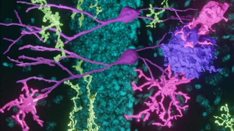
“Interestingly, SuM stimulation alone or activation of ABNs without SuM stimulation failed to restore behavioral deficits in AD mice.” Song said. “These results suggest that multi-level enhancement of ABNs — namely increasing their number, improving their developmental properties, and enhancing their activity — is required to achieve their therapeutic benefits in AD brains.” Supramammilary nucleus projections to the hippocampus (yellow), adult-born new neurons (purple), microglia (magenta), and plaques (lavender) in the Alzheimer’s mouse brain. Credit: Song Lab, UNC School of Medicine When Song and colleagues further analyzed the protein changes in the hippocampus of AD mice in response to activation of SuM-enhanced ABNs, the researchers found that several well-known protein pathways were activated inside cells.
These pathways include the ones important for synaptic plasticity of neuronal cells that allow enhanced communication among them, as well as the ones important for phagocytosis of non-neuronal microglia that allow efficient plaque clearance.
“It is striking that multi-level enhancement of ABNs through combined SuM and ABN stimulations allows such a small number of ABNs make profound functional contribution in diseased AD brains,” Song said.
“We are eager to find out the mechanisms underlying these beneficial effects mediated by activation of SuM-enhanced ABNs on AD pathology and hippocampal function. Future efforts will be needed to develop drugs that mimic these beneficial effects mediated by activation of SuM-enhanced ABNs. Ultimately, the hope is to develop first-in-class, highly targeted therapies to treat AD and related dementia.”
Funding: The National Institutes of Health funded this research through grants R01MH111773, R01MH122692, RF1AG058160, R01NS104530 to Juan Song and R21AG071229, R01GM133107 to co-author Xian Chen, PhD, professor of biochemistry and biophysics at the UNC School of Medicine. About this Alzheimer’s disease research news
Author: Mark Derewicz
Source: UNC
Contact: Mark Derewicz – UNC
Image: The image is credited to Song Lab, UNC School of Medicine
Original Research: Open access.
“ Activation of hypothalamic-enhanced adult-born neurons restores cognitive and affective function in Alzheimer’s disease ” by Juan Song et al. Cell Stem Cell
Abstract
Activation of hypothalamic-enhanced adult-born neurons restores cognitive and affective function in Alzheimer’s disease Highlights
Patterned optogenetic stimulation of SuM enhances hippocampal neurogenesis in AD
Activation of SuM-enhanced ABNs rescues memory and emotion deficits in AD
Activation of SuM-enhanced ABNs promotes hippocampal plasticity and activity in AD
Activation of SuM-enhanced ABNs increases microglia phagocytosis of plaques in AD Summary Patients with Alzheimer’s disease (AD) exhibit progressive memory loss, depression, and anxiety, accompanied by impaired adult hippocampal neurogenesis (AHN). Whether AHN can be enhanced in impaired AD brain to restore cognitive and affective function remains elusive.Here, we report that patterned optogenetic stimulation of the hypothalamic supramammillary nucleus (SuM) enhances AHN in two distinct AD mouse models, 5×FAD and 3×Tg-AD. Strikingly, the chemogenetic activation of SuM-enhanced adult-born neurons (ABNs) rescues memory and emotion deficits in these AD mice. By contrast, SuM stimulation alone or activation of ABNs without SuM modification fails to restore behavioral deficits.Furthermore, quantitative phosphoproteomics analyses reveal activation of the canonical pathways related to synaptic plasticity and microglia phagocytosis of plaques following acute chemogenetic activation of SuM-enhanced (vs. control) ABNs.Our study establishes the activity-dependent contribution of SuM-enhanced ABNs in modulating AD-related deficits and informs signaling mechanisms mediated by the activation of SuM-enhanced ABNs.Join our Newsletter I agree to have my personal information transferred to AWeber for Neuroscience Newsletter ( more information )Sign up to receive our recent neuroscience headlines and summaries sent to your email once a day, totally free.
Are you deficient in nitric oxide, the most critical molecule for cardiovascular health?
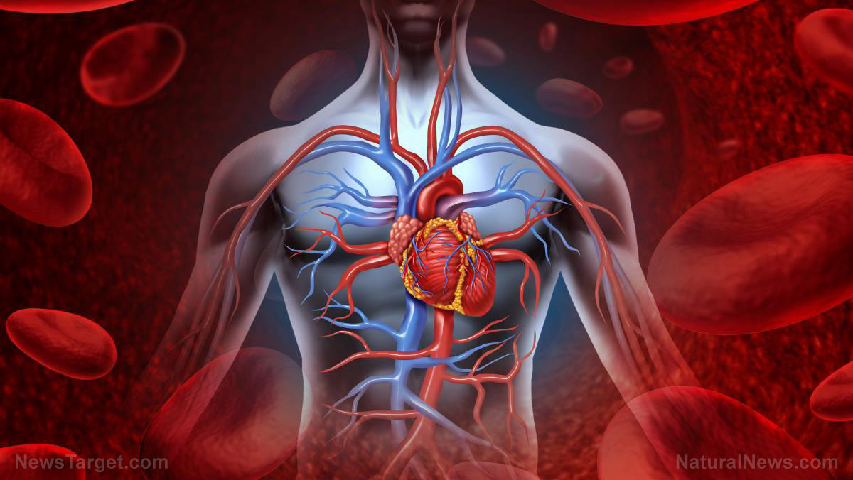
( Natural News ) One of the key components of cardiovascular health that many people overlook is a molecule called nitric oxide, or NO, which according to Nathan S. Bryan, PhD, is involved in virtually every organ system of the body.
Almost every form of cardiovascular disease begins with the loss of NO production in the body. The same is true of pretty much every chronic disease and age-related illness, whether it manifests in the kidney, the brain, the heart, or the liver.
All of these conditions have a vascular component, Bryan says. An international leader in the molecular medicine and NO biochemistry, Bryan says the common underlying factor for most chronic illnesses is a lack of oxygen-rich blood reaching certain body parts, NO being the molecule that makes it all happen.
“The lack of NO production can lead to high blood pressure, sexual dysfunction, and chronic inflammatory vascular disease leading to heart attack, stroke, or heart failure,” explains a report from The Epoch Times that cites Bryan’s work.
“The body naturally begins to produce less nitric oxide as we age but this gradual loss of NO can be sped up or slowed down based on individual lifestyle and diet.” Nitric oxide is essential for improving vasodilation, fighting inflammation, and preventing the formation of arterial plaque
Dr. Caldwell Esselstyn, the renowned director of the Heart Disease Reversal Program at the Cleveland Clinic, is another expert who says that the single most important factor impacting cardiovascular health is NO.
Its most important function throughout the body is vasodilation, which is the relaxation and widening of the inner muscles of your blood vessel. Vasodilation helps more oxygen-rich blood to travel freely throughout your body and get to where it needs to go in order to keep you healthy, fit, and strong.
Two other things Esselstyn says NO helps with is:
• Fighting inflammation by preventing arterial thickening, which can restrict blood flow, cause hypertension, and increase the heart’s workload
• Preventing the formation of plaque by reducing the “stickiness” of low-density lipoprotein (LDL) cholesterol and other elements that can build up in arteries and cause serious health problems.
Alzheimer’s disease and other forms of dementia are another class of illness for which NO can provide remedial and preventive benefits. Since the common denominator in these diseases is lack of blood flow to the brain, NO’s vasodilation, anti-inflammatory, and blood-thinning properties are a powerful weapon against dementia.
By now, you are probably wondering how you can improve your body’s NO stores, as well as how to minimize exposure to things that damage them. Here is what we know:
1) The body relies on a healthy balance of oral and gut bacterial microbiomes. Eating more probiotic-rich foods or taking probiotic supplements may help in this regard.
2) Beets, leafy green vegetables, garlic, meat, citrus fruits, nuts and seeds, and dark chocolate all contain nutritional components that help boost the body’s ability to naturally produce NO.
3) Working out, meaning exercise and rigorous activity, tells the body’s endothelial cells to produce more nitric oxide – so get moving!
“There is concrete evidence that physical activity enhances NO [nitric oxide] production,” concluded a 2021 study published in The Journal of Sports Medicine and Physical Fitness .
4) Increasing your exposure to the sun , which will also help boost your body’s vitamin D stores.
Some signs that your body may already be deficient in NO and in need of help include:
• Increased blood pressure
• Erectile dysfunction in men
• Vasculogenic sexual dysfunction in females
• Arterial plaque and inflammation
If an NO deficiency persists for too long, it can result in a heart attack or stroke, which are the leading killers of both men and women worldwide.
More related news that you can use to help support your health naturally can be found at Remedies.news .
Sources for this article include:
TheEpochTimes.com NaturalNews.com NaturalNews.com
On Nutrition: The gut-brain connection
Each type of microbe has a distinct effect on the body. (Dreamstime/TNS) If you ever thought your digestive system’s only role was to break down food, think again.
According to microbiome researcher Christopher Lowry, “what happens in the gut affects other parts of the body, including the brain.” And much of this activity is related to trillions of good and bad bacteria — collectively called our “microbiome” — that inhabit our gastrointestinal tract.
Lowry, who is currently conducting research at the Department of Integrative Physiology and Center for Neuroscience at the University of Colorado, Boulder on the relationship between stress-related disorders and the bacteria that live in our guts, was a featured speaker at a daylong workshop hosted by the Institute for Brain Potential in Cheyenne, Wyoming, titled “Understanding the Gut Brain.” 300×250 image ad “Americans are microbially deprived,” said Lowry. “Gut microbes are critical for a healthy gut and a healthy brain.”
And a diversity of good bugs appears to be the most beneficial.
That’s because each type of microbe has a distinct effect on the body. Some produce natural antibiotics to fight infections, for instance. Others enhance our body’s ability to extract energy from food. Still others produce chemicals that can actually improve our moods and calm stress. When the balance of these good bugs gets out of whack, we open ourselves to not-so-good conditions.
So how do we encourage the growth of beneficial bacteria in our guts? Spend more time in nature, Lowry suggested. That’s where we encounter a wide variety of healthful microbes.
And, consume a variety of foods that feed good bacteria. 300×250 image ad Top of the list? Plants. Health-promoting microbes thrive on fibers found in foods that sprout from the ground.
“Each plant has its own microbiome,” Lowry explained. And according to results from the American Gut Project, the largest published study to date of the human microbiome, people who ate at least 30 different types of plant foods a week had more diverse bacteria in their guts than those who ate 10 or less.
My husband and I are not vegetarians, but we do consume our fair share of fruit, vegetables, beans and grains. How hard would it be to eat 30 varieties of these foods every week?
We took the challenge. For a week, I recorded every different type of plant food we ate, no matter how much or how little. For example, our salad one night had cabbage, kale, carrots, raisins and sunflower seeds. That’s five!
The next day we had blueberries and bananas in our oatmeal, three more. Celery, onion, beans, corn, tomatoes and garlic in my stew the next day added another six to our list.
It was kind of fun … and not impossible. I found myself searching for olives to toss into egg salad. And each type of nut I ate for a snack contributed to a wider diversity of my microbiome.
The point, according to this and other researchers, is that our bodies thrive on variety. And when it comes to healthful food choices, that may be a critical key to health.
Building a better brain through music, dance and poetry
A growing body of research is probing art’s power effects on the brain. To make sense of difficult science, Michael Kofi Esson often turns to art.
When he’s struggling to understand the immune system or a rare disease, music and poetry serve as an anchor.
“It helps calm me down and actively choose what to focus on,” says Esson, a second-year student at the Medical College of Wisconsin.
Esson, who was born in Ghana, also thinks his brain is better at absorbing all that science because of the years he spent playing the trumpet and studying Afrobeat musicians like Fela Kuti .
“There has to be some kind of greater connectivity that [art] imparts on the brain,” Esson says.
That idea — that art has a measurable effect on the brain and its structure — has support from a growing number of scientific studies .
“Creativity is making new connections, new synapses,” says Ivy Ross, who is vice president of hardware design at Google and co-author of the new book, Your Brain on Art: How the Arts Transform Us .
Ross co-wrote the book with Susan Magsamen, director of the International Arts and Mind Lab at Johns Hopkins University School of Medicine. Magsamen says art’s effect on the brain is most dramatic in children.
“Children that are playing music, their brain structure actually changes and their cerebral cortex actually gets larger,” Magsamen says .
In Your Brain on Art, Magsamen and Ross describe how a person’s neural circuitry changes in response to activities like learning a new song, or a new dance step, or how to play a character on stage.
They also explain why a growing number of researchers believe these changes result in a brain that is better prepared to acquire a wide range of skills, including math and science. A brain trained to flex
Music, dance, drawing, storytelling — all of these have been a part of human cultures for tens of thousands of years. As a result, “we’re really wired for art,” Magsamen says.
And when we make art, she says, we increase the brain’s plasticity — its ability to adapt in response to new experiences.
“Children who engage in the arts are better learners,” Ross says. “Students with access to art education are five times less likely to drop out of school and four times more likely to be recognized with high achievement.”
The arts also can teach the brain skills that it’s unlikely to get in a classroom, Ross says.
“I was a dancer for like 12 years and I really think it gave me a sense of form and negative space,” she says.
Those brain circuits probably helped in her wide-ranging career, she says, which includes designing jewelry that’s part of the permanent collection at the Smithsonian.
Dancing also seems to improve mental health, Magsamen says.
“Even just 15 minutes of dance reduces stress and anxiety,” she says, noting that the activity causes the brain to release “feel-good” hormones like endorphins, serotonin and dopamine. Measuring art’s effects
The link between arts and academic achievement has been noted by educators for many years. But it’s only in the past couple of decades that technology has allowed scientists to see some of the changes in the brain that explain why.
In 2010, for example, scientists used functional magnetic resonance imaging to show that professional musicians had greater plasticity than nonmusicians in the hippocampus, an area involved in storing and retrieving information.
“The arts provide children with the kind of brain development that’s really important for building strong neural pathways,” Magsamen says, including pathways involved in focus, memory and creativity.
Esson, the medical student, may have been using some of those pathways when he found a novel way to study difficult concepts in chemistry.
“I wrote [poems] about acid-base reactions,” he says, with a laugh. “Oh my god, just so nerdy.” A failing grade for arts in school
Despite growing evidence that arts can improve performance in many other areas, activities like music and drawing have fallen out of favor in education and our culture, Ross says.
“We optimize for productivity and push the arts aside,” she says. “We thought we’d be happy. And the truth is, we’re not.”So people like Michael Kofi Esson are trying to find a balance.Now at the end of his second year of medical school, Esson spends his days on science. But sometimes late at night, he still writes poems, including one that ends with this thought about how art and the brain both create their own version of reality. Deception is art, An art the brain has mastered. Although art is a lie, It is the brain’s truth Although art is deception, it is the brain’s reality. The brain is a lie, a lie so beautiful, it is art. Esson hopes that one day he will write poems about the patients he treats. For now, though, he’s still mostly an observer.”I get to talk to them. But at the end of the day, they come for the doctor, not for me,” he says. “Once I’m actually in that position, I think I want to bring the patient into the poems.”And perhaps bring some of the poems to his patients.Copyright 2023 NPR. To see more, visit https://www.npr.org.
The brain may flush out its waste products after a mental workout
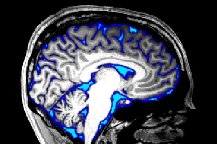
A fluid-filled network that clears waste products from the brain may be important in neurological conditions, but we know little about how it works A magnetic resonance imaging scan of an adult brain, with regions of cerebrospinal fluid overlaid in blue The brain’s “waste disposal system” may kick in after intense neural activity – and it might be possible to turn on the process intentionally.
Until recently, this system was thought to activate only during sleep, but now researchers have seen it ramping up in people after they watch flickering chequerboard patterns on a screen.
The finding provides a tantalising hint that people may be able to deliberately flush out waste products from their brain by staring at intense visual stimuli, says Laura Lewis at Boston University in Massachusetts.
Advertisement
“The real surprise was that they found it in awake people,” says Edoardo Rosario de Natale at the University of Exeter in the UK, who wasn’t involved in the work.
The brain’s waste disposal system involves cerebrospinal fluid (CSF) being pumped into the brain and leaving through a network of fine tubes called the glymphatic system , which was only discovered in 2012.
Animal research suggests the fluid flushes out waste products made by brain cells, including harmful compounds that may be involved in Alzheimer’s disease and Parkinson’s disease, such as beta-amyloid and alpha-synuclein.
Sign up to our Health Check newsletter
Get the most essential health and fitness news in your inbox every Saturday.
Sign up to newsletter
Since the glymphatic system’s discovery, there has been a surge of research aiming to understand how boosting fluid flow could help improve brain health, but much of how the system functions in people is still unclear .
Lewis’s team took advantage of a new brain-scanning technique, using existing magnetic resonance imaging machines, that highlights any CSF that has newly entered into the fourth ventricle of the brain, a cavity at the base of the head. Fluid that enters this chamber drains out through the glymphatic system.
They asked 20 volunteers to watch a screen inside the scanner that displayed a pattern known to cause high brain activity: a flickering black- and-white spiral chequerboard. The display was turned on and off at 16-second intervals for about an hour, apart from during short breaks.
Read more:
We’ve just discovered a new part of the brain’s waste disposal system
When the pattern was shown, this caused a rise in blood flow to the brain’s visual centres, as expected. When the screen went dark, blood flow reduced and CSF flow into the brain increased.
The brain-scanning technique couldn’t reveal if the fluid left through the glymphatic vessels, nor if there was a reduction of waste products within the brain. These are questions that need to be tackled next, says Rosario de Natale. “This is opening a new door.”
“It’s still an open question whether the fluid goes directly into the brain tissue or if it sloshes around in the ventricle. But we definitely think that it has an effect on fluid in the rest of the brain,” says team member Stephanie Williams , also at Boston University.
“We’re very interested now to understand the effect of these changes in fluid flow and how it intersects with brain health,” says Lewis.
Brain Health Boost Through Physical Activity, Reduced Cognitive Decline
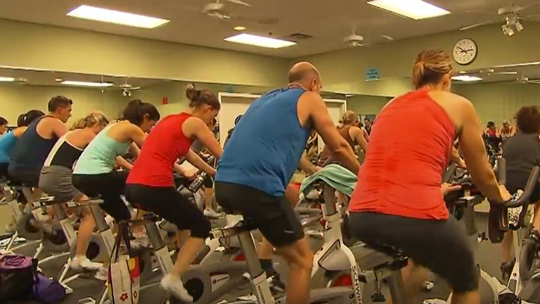
A recent release from the Center for Disease Control and Prevention (CDC ) stated that physical activity can be used to boost brain health.
UAMS shared adults need up to 150 minutes of moderate exercise a week in see benefit in the form of boosted brain health and overall quality of life.
The CDC says physical activity can significantly reduce cognitive decline including with issues such as dementia and related illnesses.
They added that this exercise can be as simple as turning on music at home and dancing. Other activities include taking your dog for a walk, parking at the back of a parking lot or getting up and marching in place when you’ve been sitting for a while.
If you participate in more vigorous physical activity it lowers to just 75 minutes per week.
Studies have shown the brain scans of those who are physically active show up younger than those who aren’t.
In addition, physical activity has been proven to help with coordination, and balance as well as improving memory and reducing anxiety and stress.
“You know we hear a lot about technology now so I’m going to make a reference I think all of us can understand. Our brain is the computer that runs it… all the operations. Every app in your body… the brain has to power those things or give them the right commands… give it the right instructions and it also has to give those commands correctly so exercising and building up those neuro pathways is one other thing that exercise does,” UAMS Chief Wellness Officer and Director of Wellness, Natalie Cannady said.
Physical activity can also help you sleep better, reduce risk of some cancers, and generally add years to your life.
Cannady did say however if you’re sensing something is off don’t solely rely on physical activity to make it better, speak with your doctor as soon as possible.
UAMS is promoting health and wellness this weekend with their “Walk with a Doc” event.
If you’re looking to kick start your physical journey with this event it will be held Saturday at 9:00 a.m. starting at the War Memorial North Lawn.
For more information on the walk click here .
Could mind games help treat teen depression? Brain imaging study shows promise
Susan Whitfield-Gabrieli, professor of psychology and director of the Biomedical Imaging Center. Credit: Ruby Wallau/Northeastern University In the midst of a devastating mental health crisis affecting thousands of American teens, Northeastern University psychology professor Susan Whitfield-Gabrieli says a non-invasive remedy for depression and anxiety offers hope.
In a brain imaging study conducted at Northeastern’s Biomedical Imaging Center, teenagers using mindfulness meditation guided by neurofeedback were able to calm a network of regions of the brain associated with depression, anxiety, ADHD and other disorders.
At the same time that region, known as the default mode network (DMN) grew less active, the central executive network (CEN) associated with goal-setting, problem-solving and sustained attention increased in function.
“Mindfulness helps with anxiety and depression,” says Whitfield-Gabrieli, whose research was published Wednesday in Molecular Psychiatry .
The study of nine people ages 17 to 19 is “proof of concept” that young people can be trained in mindfulness meditation—and that it produces results, she says.
Larger scale studies need to be done showing the efficacy of mindfulness meditation in teens and younger students, but Whitfield-Gabrieli says she and her collaborators at Massachusetts General Hospital and Columbia University are optimistic about the results from Northeastern.
So far, other therapeutic remedies for depression and anxiety, including medical and talk therapy, have a success rate of approximately 30% to 50%, Whitfield-Gabrieli says.
She says that’s not nearly enough to deal with an epidemic of adolescent mental health disorders. “We need personalized interventions that target the brain networks known to be associated with clinical symptoms,” Whitfield-Gabrieli says.
A report released in February by the Centers for Disease Control and Prevention says teen girls were experiencing record rates of sadness and more than 40% of high school survey respondents said they had felt so sad or hopeless that they could not engage in their regular activities for at least two weeks during the previous year.
“We need to catch these kids before they fall,” Whitfield-Gabrieli says. “Why not have mindfulness meditation as part of the school curriculum?”
The neurofeedback didn’t involve electrodes but had students lie in functional magnetic resonance imaging scanners that focused on specific regions of their brains.
The default mode network regions associated with depression and anxiety are activated when people worry about the future and hash over the past, Whitfield-Gabrieli says, adding that she calls engaging this area of the brain “mental time travel.”
When the default mode network regions are activated, the central executive network regions associated with the ability to manage tasks and regulate moods show less activity, Whitfield-Gabrieli says.
The activity in the different areas of the brain rises and falls in a type of dance or synchrony that is more pronounced the more psychopathology is present, she says.
The goal of the researchers was to use fMRI signals to moderate the activity, and it worked, Whitfield-Gabrieli says.
“As soon as the person starts doing the mindfulness meditation, the DMN drops,” she says.
To make the exercise even more engaging for the teens, the researchers converted it into a game.
When the participant got the DMN to reduce activity, they’d get a signal to move a ball up towards a target circle. But if that region of the brain started getting more active, the ball would drop.
“They are literally just moving the ball with their mind. They love it,” Whitfield-Gabrieli says.
“Mindfulness meditation forces you to attend to the present moment. You are no longer caught in the past or the future,” she says.
There’s no reason not to incorporate mindfulness meditation and guided breathwork into school curricula now, Whitfield-Gabrieli says.
She says eventually she would like to see neurofeedback work with other biomarkers such as heart rate to develop interventions for when people fall into vulnerable states of repetitive negative thinking.
In the near future, a smart phone might be able to pick up on the signals and request the user to “do mindfulness meditation now,” exercise or remember to take an antidepressant, she says.
“People are realizing more and more the amazing effective and global impact mindfulness meditation can have.”
More information: Jiahe Zhang et al, Reducing default mode network connectivity with mindfulness-based fMRI neurofeedback: a pilot study among adolescents with affective disorder history, Molecular Psychiatry (2023). DOI: 10.1038/s41380-023-02032-z
Provided by Northeastern University

The Power Within by Corey Daniels book available for only $2.99
Mind has both conscious and subconscious halves. These are likened to a driver and the truck he drives. The driver plans the destination and observes road conditions, while the truck provides motive power. Your subconscious mind is like the truck and it only goes in the direction in which is a steered. This can be the road or off a cliff. Likewise, the consciousness paints a picture of what the world is and what your goals are and the subconscious acts on them through emotion, physical response and energy, whether these are correct, rational images or false negative ones. The subconscious is also like an emotional reservoir which your body and mind draw responses from to external stimuli.
The Power Within lays out a method of programming your subconscious and tapping into the Holy Spirit, God voice or what the Greeks called the daimon (godman).