Nature Knows and Psionic Success
God provides
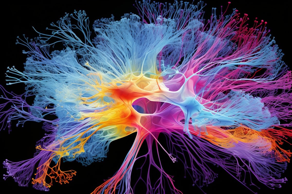
Cellular Map of Entire Brain Reveals What Makes Us Human
Summary: A groundbreaking suite of 21 papers has unveiled a momentous leap in our understanding of the brain, spotlighting the intricate cellular composition of human and primate brains through a consortium led by the BRAIN Initiative.
Utilizing innovative single-cell transcriptomics, researchers illuminated a stunning array of over 3,000 different brain cells and their distinctive functionalities. This extensive research not only dives into the distinctiveness of the human brain but also pioneers a suite of scalable techniques that offer an unparalleled, detailed organization view of the brain.
This pivotal moment in neuroscience sets a promising stage for the next phase in cellular census efforts, propelling towards a more profound understanding of the brain’s complexity and functionality.
Key Facts:
> Vast Cellular Diversity: Scientists discovered an astonishing array of more than 3,000 different kinds of brain cells through a technique called single-cell transcriptomics.
Global Collaboration: This massive research effort involved hundreds of scientists worldwide and is funded by the National Institutes of Health’s BRAIN Initiative, aiming to explore and catalog the complex cellular makeup of primate brains, including humans.
Comparative Study: The research also undertook a comparative study between human brains and those of our closest ape relatives, revealing both shared cellular architectures and significant variances in gene utilization within neuron connections and circuit formations.
Source: Allen Institute
Scientists have just unveiled a massive effort to understand our own brains and those of our closest primate relatives.
In a suite of 21 papers published today in the journals Science (12) , Science Advances (8) , and Science Translational Medicine (1) , a large consortium of researchers shares new knowledge about the cells that make up our brains and the brains of other primates. It’s a huge leap from previously published work, with studies and data that reveal new insights about our nervous systems’ cellular makeup across many regions of the brain and what is distinctive about the human brain.
The research consortium is a concerted effort to understand the human brain and its modular, functional nature. It was brought together and is funded by the National Institutes of Health’s Brain Research Through Advancing Innovative Neurotechnologies ® (BRAIN) Initiative.
Hundreds of scientists from around the world worked together to complete a range of studies exploring the cellular makeup of the human brain and those of other primates, and to demonstrate how a transformative new suite of scalable techniques can be used to study the detailed organization of the human brain at unprecedented resolution.
Understanding our brain at the cellular level is key to understanding how our brains function and who we are as a species, as well as more accurately pinpointing the cellular roots of brain diseases and disorders—knowledge that could ultimately lead to better treatments for those diseases.
Scientists at the Allen Institute for Brain Science, a division of the Allen Institute, led five of these studies and made significant contributions to three others, including a study that greatly expands on existing knowledge about the number of types of cells in the adult human brain.
Scientists at Karolinska Institute and the Allen Institute studied the genes switched on in individual brain cells, a technique known as single-cell transcriptomics, revealing an astonishing diversity of cell types: we have more than 3,000 different kinds of brain cells.
“I view this as a pivotal moment in neuroscience, where new technologies are now allowing us to understand the very detailed cellular organization of the human brain and of other primate brains,” said Ed Lein, Ph.D., Senior Investigator at the Allen Institute for Brain Science, who led several of the newly published studies.
“At its core, this body of work is a triumph of molecular biology: Differential gene usage can be used to define cell types, and the tools of genomics could be used to create the first drafts of high-resolution, annotated maps of the cells that make up the entire human brain.”
The studies also tackle a range of important questions such as: How different are individual people’s brains at the cellular level? How different are our brains from those of our closest ape relatives? How many kinds of brain cells do we have? What are the properties of these cells? How do these cells emerge and mature in development?
Building off previous work mapping brain cell types in high resolution in single regions of the human cortex, the outermost shell of the brain, the newly published package expands those studies to dozens up to a hundred regions across the entire brain.
Where the single region studies found over 100 different brain cell types, the newly released data shows thousands of different kinds of brain cells across the entire brain. For many parts of the brain, that complexity and variety had never before been described.
These studies are part of the NIH’s BRAIN Initiative Cell Census Network, or BICCN, a five-year funding program that was launched in 2017 to create a catalog of brain cell types. This body of work demonstrated the scalability of cutting edge cellular and molecular approaches to tackle the challenges of size and complexity of the human brain, and has set the stage for the next phase of this cell census effort.
This next phase, part of which is underway at the Allen Institute, will build much more comprehensive atlases of human and other primate brains through the BRAIN Initiative’s Cell Atlas Network, or BICAN.
“The present suite of studies represents a landmark achievement that continues to build an important bridge toward illuminating the complexity of the human brain at the cellular level,” said Dr. John Ngai, Director of the NIH BRAIN Initiative.
“The scientific collaborations forged through BICCN, and continuing in the next phase in BICAN, are propelling the field forward at an exponential pace; the progress – and possibilities – have been simply breathtaking.”
The human studies used postmortem tissue from people who had donated their brains to science, as well as healthy living tissue donated from patients who had undergone brain surgery and donated tissue to research.
The data from the newly released studies will also feed into the Human Cell Atlas, an international effort that […]
Read more at neurosciencenews.com

The Power Within by Corey Daniels book available for only $2.99
Mind has both conscious and subconscious halves. These are likened to a driver and the truck he drives. The driver plans the destination and observes road conditions, while the truck provides motive power. Your subconscious mind is like the truck and it only goes in the direction in which is a steered. This can be the road or off a cliff. Likewise, the consciousness paints a picture of what the world is and what your goals are and the subconscious acts on them through emotion, physical response and energy, whether these are correct, rational images or false negative ones. The subconscious is also like an emotional reservoir which your body and mind draw responses from to external stimuli.
The Power Within lays out a method of programming your subconscious and tapping into the Holy Spirit, God voice or what the Greeks called the daimon (godman).