Nature Knows and Psionic Success
God provides
Battle of the Dendrites: How Neurons Compete To Cut Connections
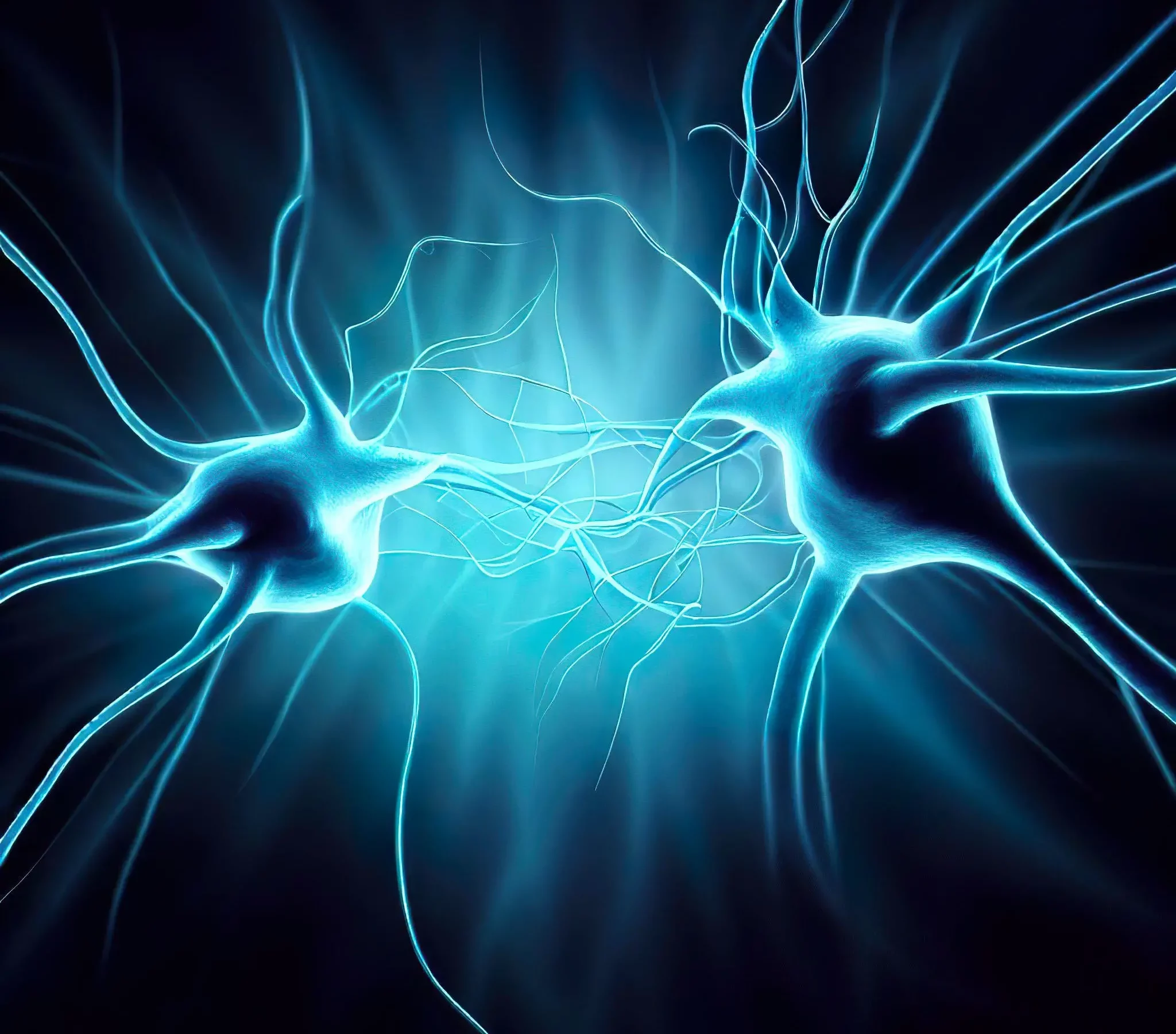
Researchers at Kyushu University discovered the chemical pathways that regulate synaptic pruning, a crucial phase in brain development where excessive and incorrect neuronal connections are eliminated. The team found that in the presence of neurotransmitter signaling, the receiving dendrite is protected while other dendrites of the same neuron are set on a path to be pruned, a mechanism that helps refine neural networks and contribute to proper brain maturation. Scientists elucidate the process through which synapses compete with each other, and describe how during development, weak and noisy synapses are eliminated during development.
Scientists from Kyushu University have uncovered the mechanisms underlying a crucial but often overlooked stage in brain development known as synaptic pruning.
The research team used mouse mitral cells, a kind of neuron in the olfactory system, for their study. They discovered that when neurons accept a neurotransmitter signal, the recipient dendrite is shielded via a sequence of chemical pathways. Simultaneously, the depolarization triggers other dendrites from the identical cell to follow a separate pathway that promotes pruning. The findings were recently published in the journal Developmental Cell .
How neurons connect and remodel themselves is a fundamental question in neurobiology. The key concept behind proper networking is in neurons forming and strengthening connection with other neurons while pruning excessive and incorrect ones.
“A common phrase in neural circuit remodeling is ‘fire together wire together’ and ‘out of sync, lose your link.’ The former describes how neurons that pass signals between each other tend to strengthen connections, whereas the latter explains that without said signaling that connection diminishes,” explains Professor Takeshi Imai from Kyushu University’s Faculty of Medical Sciences, who led the study. “It’s a refining process that is fundamental for proper brain maturation.”
Olfactory bulb of mouse two days after birth with fluorescence indicating signaling. The video shows that glomeruli, the signaling way station in the olfactory bulb, spontaneously send out signals. This spontaneous signaling will eventually lead to proper networking and pruning of mitral cells. The video was imaged ex vivo using two-photon microscopy. Credit: Kyushu University/Imai Lab
Over the decades, researchers—including Prof Imai—have explored the fundamental process of how neurons form and strengthen their connections. However, there had been one major gap in the process that few people were examining: how the connections are eliminated.
“The elimination of neuronal connections, what we call pruning, was something everybody in the field knew about and observed. But if you look at the literature, there was a lack of study on the exact mechanism that drove the process,” explains first author Satoshi Fujimoto.
Elimination of connections happens everywhere in the nervous system, for example in neuromuscular junctions, the neurons that send signals to your muscles to move. At first, the muscle fibers receive inputs from many motor neurons. As you grow, these connections are finetuned, where some are strengthened, and others are eliminated, until just one neuron connects to one muscle fiber. It is why you have awkward motor control and coordination at an early age.
In early development, neurons called mitral cells grow multiple branches to connect with multiple glomeruli. Like a bonsai, as development progresses branches get strengthened and pruned. But while researchers investigated closely the mechanism of branch strengthening, how pruning was induced remained under-studied. Kyushu University researchers found that when mitral cells receive the neurotransmitter glutamate, the subsequent signal triggers local suppression of RhoA, protecting that dendrite. At the same time, the depolarization activates the pruning machinery—controlled by RhoA—in dendrites that did not receive the glutamate input. The winner dendrite takes all. Credit: Kyushu University/Imai Lab
“We decided to investigate what exactly happens in neurons during remodeling, so, we looked into using mouse mitral cells, a type of cell housed in the olfactory bulb, the brain center involved in our sense of smell. In adults, mitral cells have a single connection to a signaling waystation called the glomerulus. But in early development mitral cells send branches into many glomeruli,” states Fujimoto. “As time progresses, these branches get pruned to leave a single strong connection. In the end, the mitral cells can sniff out only a specific type of smell.”
First, the team found that spontaneous waves of the neurotransmitter glutamate in the olfactory bulb facilitate dendrite pruning. The team then focused on the mitral cell’s inner signaling pathways. What they found was a unique protection/punishment machinery that would strengthen certain connections and kick off the pruning of others.
“We found that in the mitral cells it was the signaling from glutamate that was essential for pruning. When glutamate binds to its receptor NMDAR in a dendrite, it suppresses the pruning machinery molecule called RhoA,” continues Fujimoto. “This ‘save-me’ signal is important to protect it from pruning.”
From the moment mice are born, their mitral cells extend multiple dendrites into multiple glomeruli. They form branches and excitatory synapses in the glomerulus at around day three after birth. By day six, they form single dendrites through selective pruning. This makes it possible to receive information from only one type of olfactory receptor (odor sensor), which is the basis of odor discrimination. Credit: Kyushu University/Imai Lab
Upon the glutamate input, the mitral cell also depolarizes and fires a signal. The team also found that depolarization triggers the activation of RhoA in other dendrites of the same cell, and kicking off the pruning process. Simply put, the dendrite that receives the direct glutamate signal is protected, while the other dendrites get pruned.
“This ‘punishment’ signal for synapse elimination only acts on non-protected synapses, and it explains how only a strong connection becomes the winner and all the others mediating weak and noisy inputs become the losers,” Imai explains.
The team’s findings reveal new information about an over-looked but critical phase in neural development.
“Proper pruning of neuronal connections is just as important as the strengthening of the network. If it goes awry in either direction it can lead to different kinds of neurophysiological disorders. Too few connections have been linked to schizophrenia, whereas too many connections have been found in people with autism spectrum disorder, for example.” says Imai. “To […]
Do Brain-Boosting Supplements Actually Work Or Are They Total B.S.?
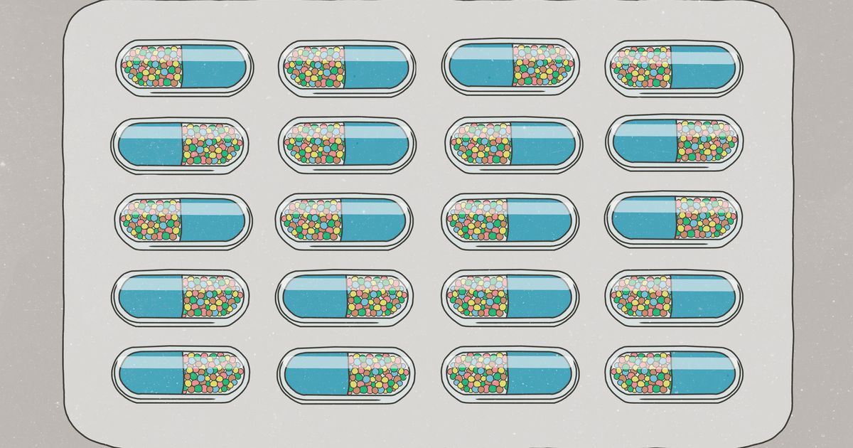
Can a supplement really make you smarter?
A quick online search for “mental clarity supplements” will turn up tons of products, such as the Neuriva Brain Health supplement , Neurohacker Collective’s “Qualia Mind,” WTHN’s “Fully Charged,” and Bulletproof’s “Unfair Advantage, ” just to name a few. These supplements claim to improve focus, sharpen thinking, boost productivity and fight fatigue.
It all sounds like something straight out of a gimmicky infomercial ― just another quick-fix product to buy to improve your health. But as it turns out, there is some science that supports the brain-boosting power of some (yet not all) of the ingredients in mental clarity supplements. And, of course, there’s also a catch (there’s always a catch).
Here’s what you should know before you purchase them, according to experts: Which Brain Supplement Ingredients Work And Which Need More Research
The more scientific classification of these supplements and their ingredients is called nootropics. The term, coined by psychologist Corneliu E. Giurgea in 1972 , describes natural or man-made substances designed to support cognitive functioning and improve mental performance. Other terms you may have heard for them could include “smart drugs” or “cognitive enhancers.” They range from familiar substances — such as caffeine or turmeric — to complex blends of many different herbs and other ingredients sold online.
Now, for the million-dollar question: Do they work? Well, yes and no. The reason for this ambiguous answer? The research on brain-boosting supplements is ambiguous itself. While some of the ingredients in brain-boosting supplements have shown to be beneficial, the studies have been inconsistent, explained Dr. Michael Genovese , a clinical psychiatrist and chief medical officer of Acadia Healthcare.
Plus, “unlike other medications, the [Food and Drug Administration] does not regulate the distribution of so-called ‘nootropics,’ so there is no way to tell with certainty if the label on the bottle is what is really in the supplement,” he added.
Dr. David J. Puder , medical director of the behavioural health outpatient program at the Loma Linda University, puts it a little more bluntly: “The people making these supplements are not doing randomised controlled trials to determine if they really work,” he said. “They mostly use data about what might work, throw it together and add a bunch of excellent marketing.”
So, with that said — and a healthy dose of skepticism — let’s dive into what the research says about a few common ingredients used in mental clarity supplements: Caffeine
Surprise, surprise. Many mental clarity supplements contain a very familiar ingredient, which is good old-fashioned caffeine. And for good reason: “It is commonly understood that caffeine reduces drowsiness and increases alertness and psychomotor performance,” Puder said. Plus, research shows caffeine use may have a protective effect on memory . Omega-3 Fatty Acids
Another nootropic you’ve likely heard of before is omega-3 fatty acids, namely EPA and DHA, which are critical for proper brain function and development. Several studies have shown that taking fish oil supplements (which are high in EPA and DHA) may help improve memory in people with mild cognitive impairment, and they are also recommended to help people with depression. One caveat: Research did not find improvements in healthy people’s cognitive function after taking fish oil supplements. Ashwagandha
A highly revered herb in the Ayurvedic tradition (an ancient Indian healing system), ashwagandha is known as an adaptogen, which means it may help protect the body from the effects of stress . Research has also found ashwagandha may help enhance focus, mental stamina and can even be useful for people with neurodegenerative diseases such as Parkinson’s, Huntington’s and Alzheimer’s diseases. Rhodiola
A perennial flowering plant, rhodiola is a nootropic that studies have shown can decrease fatigue and increase the capacity for mental work, said Shari Auth, a holistic health practitioner in New York and co-founder of WTHN , a modern acupuncture studio with a line of herbal supplements. People with diagnoses like ADHD and anxiety disorders may also benefit from rhodiola, since it helps the mind adapt to stressors, Genovese added. Bacopa Monnieri
Another Ayurvedic herb, Bacopa has traditionally been used to enhance brain function. “As a nootropic, it acts as a ‘brain tonic’ to enhance memory, cognitive function and reduction of cortisol levels, which alleviates stress in the body,” Auth said. Several scientific studies back this up, although it may require long-term use to reap the results. Ginkgo Biloba
Derived from the leaves of a tree called Ginkgo biloba, this supplement has been shown to improve memory and mental processing in healthy older adults when taken daily for six weeks. Taking Ginkgo biloba before a highly stressful task can help lower stress-related high blood pressure, as well as high levels of cortisol, a type of stress hormone.
Genovese added that he’s heard anecdotal evidence that people have seen improvements in loved ones with Alzheimer’s after they supplemented with Ginkgo balboa. However, that’s yet to be determined through research. What To Keep In Mind Before You Buy Brain Supplements
It’s important to note that simply taking one or two of these nootropic agents often results in no significant net change, explained Dr. Alex Dimitriu, founder of Menlo Park Psychiatry & Sleep Medicine .
Instead, researchers have found that a concoction of some of the most researched and validated supplements, with overlapping benefits, is a better approach. “The hope is that if you have enough ingredients with overlapping benefits, taken together, they may be more effective than taken individually,” Dimitriu said.
That’s why you’ll see long lists of ingredients of so many of the popular “mental clarity” supplements on the market. One popular product called Neurohacker Collective’s Qualia Mind , for instance, contains a laundry list of ingredients that contain six nootropics, along with other vitamins and adaptogens.
Most experts we chatted with said trying the supplement won’t be harmful. (Always check with your doctor first before trying something, though.) But if you’re gawking at the prices, don’t sweat it — you really don’t need a supplement to feel “smarter.”
Instead, focus on the basic […]
Tips to improve brain health with better screen habits
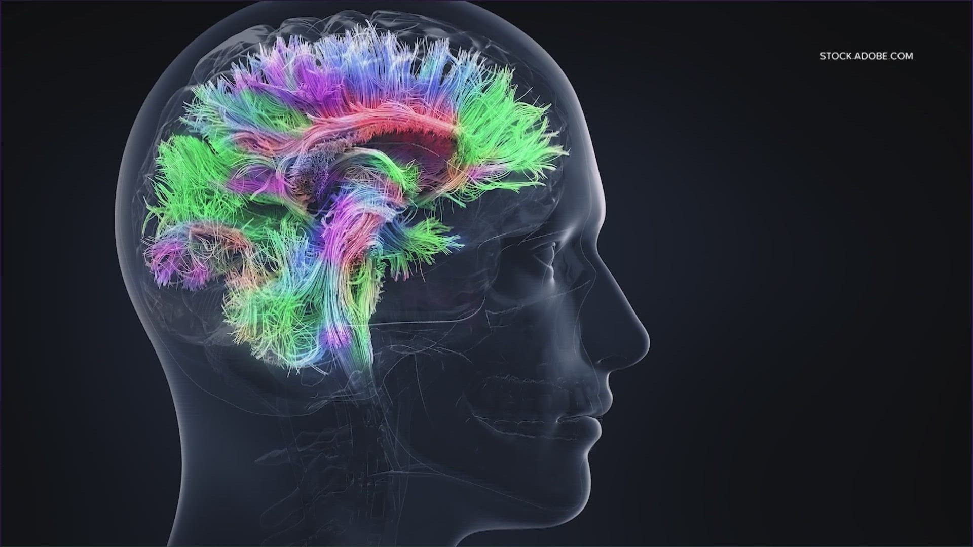
Excessive screen time and social media consumption has been linked to anxiety, depression and other brain-based issues. AUSTIN, Texas — June is recognized as Brain Health Awareness Month, and everyday activities like social media consumption and excessive screen time have been linked to brain-based issues.
Alex Fink , founder and CEO of Public Benefit Corporation Otherweb , says multiple studies link excessive media consumption to an increased risk of depression, anxiety, loneliness and insomnia. Whether it’s endlessly scrolling through social media posts or staring into bright screens, media overload is hurting the brain and mental health.
“Over time, you start talking to people, they start talking about things like gaps in their day, They start talking about spending excessive amounts of time and not knowing why or not remembering what it is that they did during this time. You see people becoming more irritable, more nervous, more agitated over time. I think these are all the effects of addiction, whether it’s addiction to a drug or to social media, which acts as a kind of a drug,” said Fink. Related Articles
Fink says there are solutions to improving screen health, including being mindful of the content you’re interacting with. Also, plan how much time you want to spend consuming content that may not be necessary for your family or work life.
“Try to divide the time in your head. Like if you’re going to spend 2 hours a day consuming content, how much of that should be books? How much of those should be news? How much of that should be checking on your friends and what they’re up to? If you have this plan in your head and you try to stick to it and you try to control where you’re consuming content, what it is that you’re consuming, next, chances are your diet will end up being much better than it would otherwise have been,” stated Fink.
For children to beat screen habits, Fink said the solution is pretty simple. Sit down with them and talk about how their media programs work and what they are trying to do.
“What are you trying to get out of it? How do you avoid the pitfalls of the program essentially taking control? Because we have to remember our kids don’t remember the way things used to be. They don’t have a baseline. They don’t feel that something has changed. This is the first thing that they encounter and this thing incorporates the knowledge of many ideas that have been working on this, whereas our kids are kind of just one person with one person’s experience, so we have to arm them before letting them go into this battle,” said Fink.
Another solution is exercise. Fink says exercising every day has been proven to be the most effective remedy against any brain ailment, disease or discomfort.
“It’s the best solution to depression. The best solution to attention deficit disorder. Just run or bike for 20 minutes a day. Beyond that, I think just being mindful and thinking about it is already a step in the right direction,” said Fink.
Fink’s focus has not just been about helping people control their screen time, but on helping people control the quality of the content they consume during this time. The CEO believes there is a pull on all content, on newspaper editors, on social media, on everyone to just maximize clicks and views and their viewers.
“Obviously the thing that makes you click the most is not the most important thing for you to know right now. So our company, would try to improve the quality of content that people consume and try to give people the tools to select content better. I’m not advocating that people use this specifically, but try to seek out tools that help you control what it is that you’re putting into your brain,” said Fink.
Decoding Neurological Mysteries: How Blood Triggers Brain Disease
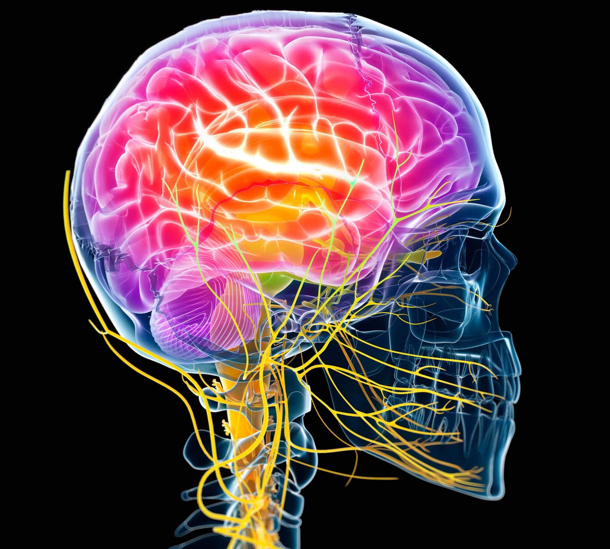
A new study from the Gladstone Institutes has identified a blood protein called fibrin as the key factor that transforms beneficial immune cells in the brain, known as microglia, into harmful ones, which contribute to diseases like Alzheimer’s and multiple sclerosis. New insights into how blood makes the brain’s immune cells toxic point to new therapeutic strategies for Alzheimer’s disease and multiple sclerosis.
In individuals suffering from neurological disorders such as Alzheimer’s and Multiple Sclerosis, beneficial microglia, the immune cells within the brain, turn harmful to neurons. This detrimental shift contributes to cognitive dysfunction and impaired motor skills. Additionally, these harmful immune cells might play a role in cognitive decline associated with aging in individuals who are not suffering from dementia.
For a while, researchers have been diligently working to comprehend what precisely prompts these beneficial microglia to become harmful, and their specific role in disease progression. If they could identify what makes microglia toxic, they could find new ways to treat neurological diseases.
Now, researchers at Gladstone Institutes led by Senior Investigator Katerina Akassoglou, Ph.D., showed that exposure to blood leaking into the brain turns on harmful genes in microglia, transforming them into toxic cells that can destroy neurons.
The scientists discovered that a blood protein called fibrin—which normally aids blood clotting—is responsible for turning on the detrimental genes in microglia, both in Alzheimer’s disease and multiple sclerosis. The findings, published in the journal Nature Immunology , suggest that counteracting the blood toxicity caused by fibrin can protect the brain from harmful inflammation and loss of neurons in neurological diseases.
“Our study answers, for the first time in a comprehensive way, how blood that leaks into the brain hijacks the brain’s immune system to cause toxic effects in brain diseases,” says Akassoglou, who is also director of the Center for Neurovascular Brain Immunology at Gladstone and a professor of neurology at UC San Francisco (UCSF) . “Knowing how blood affects the brain could help us develop innovative treatments for neurological diseases.”
Katerina Akassoglou (left) and Andrew Mendiola (right) show how blood makes the brain’s immune cells toxic, pointing to new treatments for Alzheimer’s disease and multiple sclerosis. Credit: Michael Short/Gladstone Institutes Specific Effects of Blood Proteins
Individuals with neurological diseases like Alzheimer’s disease and multiple sclerosis have abnormalities within the vast network of blood vessels in their brain, which allow blood proteins to seep into brain areas responsible for cognitive and motor functions. Blood leaks in the brain occur early and correlate with worse prognosis in many of these diseases.
To understand which proteins in the blood affect gene and protein changes in immune cells, Akassoglou and her team took a systematic approach to determine how losing key blood proteins—such as albumin, complement, and fibrin—would affect immune cells in mice.
They analyzed the effect of the blood proteins with a suite of advanced molecular and computational technologies in collaboration with Nevan Krogan, Ph.D., senior investigator at Gladstone and director of the Quantitative Biosciences Institute at UCSF, and Alex Pico, Ph.D., research investigator and director of the Bioinformatics Core at Gladstone.
In the new study, the researchers found that different blood proteins activate distinct molecular processes in microglia. What’s more, they identified that fibrin is responsible for driving unique gene and protein activities that make microglia toxic to neurons. The other blood proteins tested were not mainly responsible for these toxic effects.
“We combined cutting-edge tools to capture a broad view of all the microglia processes triggered by distinct blood proteins,” says Andrew Mendiola, Ph.D., a scientist in Akassoglou’s lab and first author of the study. “Fibrin stood out, as it triggered a dramatic gene response in microglia, which mirrored gene signatures identified in chronic neurological diseases such as Alzheimer’s disease.”
In prior research, Akassoglou and her team discovered that fibrin can activate microglia and promote cognitive impairment in mice. Indeed, the researchers were able to narrow down fibrin’s bad influence to a specific inflammatory region of the protein. This region does not impact fibrin’s critical role in blood clotting. In the new study, the team showed that removing that inflammatory region reduced fibrin’s ability to turn on toxic genes in microglia, and restored the protective functions of these immune cells. Implications for Neurological Diseases and Therapies
To assess whether their findings are relevant to disease, the researchers used a technique they developed to identify toxic gene activities in cells in mouse models of Alzheimer’s disease and multiple sclerosis. In both types of models, fibrin-activated microglia genes are involved in neurodegeneration and oxidative stress, processes that have been linked to both Alzheimer’s disease and multiple sclerosis.
“We think that, across neurological diseases, fibrin deposits at sites of blood leaks might drive toxic immune responses,” Mendiola says. “Identifying approaches to selectively inhibit these toxic responses could be a game changer for treating disease.”
Akassoglou’s lab has already developed one such drug , a therapeutic monoclonal antibody against fibrin’s inflammatory domain. The antibody blocks the detrimental effects of fibrin without adverse effects on blood clotting, and protects from multiple sclerosis and Alzheimer’s disease in mice. A humanized version of this first-in-class fibrin immunotherapy has now started Phase 1 safety clinical trials .
“Neutralizing blood toxicity could protect the brain from harmful inflammation and restore neuronal connections required for cognitive functions,” says Akassoglou. “By targeting fibrin, we can block toxic microglia cells without affecting their protective functions in the brain.”
The study generated a large amount of molecular data that is now freely available for other researchers to use. The open-access atlas of how the blood affects the brain could be further analyzed to reveal other functions of blood proteins and support the discovery of new drugs and biomarkers.
“These exciting findings change the way we think about blood proteins, from secondary bystanders to primary drivers of harm in the brain,” says Lennart Mucke, MD, director of the Gladstone Institute of Neurological Disease. “The mechanisms identified in this study could be at work in a range of neurological conditions involving blood leaks in the brain, including neurodegenerative disorders, autoimmune diseases, stroke, and traumatic brain injury. Therefore, they […]
New Report Highlights Recommendations for Achieving Better Brain Health for All
AARP and the Global Council on Brain Health recognize barriers that impact some groups more than others and offer solutions to encourage cognitive wellness for all
WASHINGTON —Brain health is influenced by many factors, including economic and social factors such as income and financial security, housing conditions, environment, and access to nutritious food and exercise. A new report from AARP and the Global Council on Brain Health (GCBH), “ Building Better Brain Health for All People: GCBH Recommendations on Removing Barriers and Improving Opportunities Around the World ,” takes these into account and offers recommendations for achieving greater brain health.
“If we want to improve brain health for all, we have to pay more attention to the needs of those at greatest risk of poor health and address social conditions that stand in the way,” said Sarah Lenz Lock, Senior Vice President of Policy and Brain Health at AARP and Executive Director of the Global Council on Brain Health. “Cognitive decline is not inevitable, and everyone should have the opportunity to experience better brain health as they age.”
The report highlights the ‘ Six Pillars of Brain Health ’ that can support the ability of individuals to positively affect their brain health. The pillars include exercise, intellectual stimulation, sleep quality, stress management, social engagement, and nutrition. While these may sound simple, many obstacles can prevent people from incorporating the six pillars into their everyday lives.
Recommendations in the report for addressing barriers to brain health are informed by the latest scientific findings and by lived experience of people with cognitive issues. These include: For individuals : Be an active participant in your health care and seek providers you can trust to listen and understand your cultural values. Make healthy choices whenever possible and try to incorporate the Six Pillars of Brain Health into your lifestyle.
For health care providers: Prioritize prevention and establish brain health screening as an important element in check-ups of aging adults and others at risk. Ensure family caregivers have the information they need to provide the most effective care and include them in consultations as appropriate.
For policymakers: Establish public policies and practices to promote greater awareness and early identification of cognitive and mental health issues, including diverse voices in the policymaking process. Facilitate healthy lifestyles enabling people to proactively promote and sustain their brain health across their lifespans.
For communities: Promote community engagement to raise awareness of brain health and debunk misconceptions. As community leaders, employers can take the lead in creating incentives to encourage healthy behavior. Include community members in decision-making and incorporate diverse perspectives, expertise, and feedback in all education, outreach, and communications initiatives.
“Better brain health enables people’s minds to flourish so they can lead more rewarding lives,” said Lock. “Successful policies and strategies to promote brain health must integrate the many elements that influence cognitive wellness throughout life.”
Read the full report here and view the infographic .
# # #
About AARP
AARP is the nation’s largest nonprofit, nonpartisan organization dedicated to empowering people 50 and older to choose how they live as they age. With a nationwide presence, AARP strengthens communities and advocates for what matters most to the more than 100 million Americans 50-plus and their families: health security, financial stability and personal fulfillment. AARP also produces the nation’s largest circulation publications: AARP The Magazine and AARP Bulletin. To learn more, visit www.aarp.org/about-aarp/ , www.aarp.org/español or follow @AARP , @AARPenEspañol and @AARPadvocates on social media.
For further information: Emily Pickren, epickren@aarp.org, 202-431-7752
Study: Consuming walnuts found to reduce the negative effects of stress

Walnuts are known for the many health benefits they offer , which include supporting a healthy gut, lowering the risk of Type 2 diabetes and fighting certain types of cancer.
But perhaps their best-known contribution to human health is their ability to improve brain function and reduce the risk of cognitive decline in older adults. This benefit, according to studies, stems from their abundance of plant-based omega-3 fatty acids and polyphenols, which can reduce brain inflammation. Chronic brain inflammation is believed to be a key driver of neurodegenerative disorders like Alzheimer’s disease and dementia.
A clinical trial conducted by researchers from the University of South Australia (UniSA) has found that walnuts also provide mental health benefits. Daily walnut consumption in a small group of undergraduate students was shown to effectively reduce stress and depression and counteract the negative effects of stress on the gut and metabolic health. How stress impacts mood, gut microbiota and metabolic markers
Stress can affect all systems of the body and is known to cause a number of symptoms . Among these, dramatic changes in mood and appetite, pain, depression and stomach discomfort are the most common.
Your body produces high amounts of cortisol when you are under a lot of stress. Cortisol is the primary stress hormone released as part of the body’s fight-or-flight response to stimulate the liver to produce more glucose and facilitate the conversion of nutrients into energy. These are meant to naturally boost energy levels to enable the body to better respond to the cause of stress.
But having persistently high cortisol levels, as in the case of chronic stress, has negative consequences, particularly for mental health. Studies show that an elevated cortisol response is associated with acute and severe forms of major depressive disorder . Abnormal cortisol levels can also lead to the development of Cushing syndrome , a condition that causes weight gain, high blood pressure and mood changes.
Aside from mood, stress also has a negative impact on the gut and commensal bacteria. Research has found that during times of stress, distress signals reach the gut via a pathway mediated by immune cells . This event triggers inflammation, which favors the growth of certain gut bacteria that could become pathogenic when allowed to flourish.
A stress-induced shift in gut microbial composition can also cause an imbalance (dysbiosis) and increase the permeability of the gut barrier, resulting in a “leaky gut” that allows potentially harmful bacteria to seep into circulation. Gut dysbiosis and decreased diversity of gut commensal bacteria have been linked to chronic conditions like obesity and Type 2 diabetes, and neurodegenerative disorders like Alzheimer’s and Parkinson’s in the elderly.
Multiple studies have also reported a link between stress and metabolic disorders. In fact, chronic stress is considered an important risk factor for metabolic syndrome , a cluster of conditions that occur together and increase a person’s risk of heart disease, stroke and diabetes. Other risk factors used to diagnose metabolic syndrome include excess belly fat, high blood pressure, high triglyceride levels, low high-density lipoprotein cholesterol (good cholesterol) and high fasting blood sugar.
According to studies, stress has a direct influence on these metabolic markers. For instance, a report published in the journal Medicine found that chronic stress can increase blood triglyceride and low-density lipoprotein (bad cholesterol) levels and decrease high-density lipoprotein. High cortisol levels have also been linked to abdominal obesity as it can trigger uncontrollable eating .
And because cortisol promotes the production of glucose, chronic stress can keep your blood sugar elevated and put you at risk of developing Type 2 diabetes. This is why effective stress management is very important, especially for people constantly exposed to various stressors in their daily lives. Having three of the five risk factors mentioned above is enough to be diagnosed with metabolic syndrome. Walnuts can help students deal with academic stress
Academic stress is a chronic stressor that negatively affects the mental health of university students. According to the National Alliance on Mental Illness, about 75 percent of mental health disorders develop prior to the age of 24 . Because undergraduate students are continually subjected to academic stress, they are particularly prone to depression. A 2022 study corroborates this, with survey results showing an overall 135 percent increase in depression and a 110 percent increase in anxiety among college students across the U.S. from 2013 to 2021.
As multiple studies have pointed out, chronic stress leads to the dysregulation of the body’s stress response system, which results in chronically high levels of cortisol. Excessive amounts of cortisol have been shown to impair learning and memory and disrupt the mesolimbic dopaminergic system, a key component of the brain’s reward pathways. The resulting decrease in reward sensitivity is what ultimately leads to feelings of depression.
But recent investigations into dietary interventions that could help prevent stress-induced depression have found that walnut consumption exerts positive effects on both cognition and mental health. These benefits appear to be linked to walnuts’ abundance of brain nutrients, such as omega-3 fatty acids , a -linolenic acid and tryptophan, and compounds that support cognition, such as melatonin, polyphenols, folate and vitamin E.
Thanks to these brain-supporting components as well as walnuts’ fiber content, regular walnut intake has also been reported to promote gut microbial diversity. Because of the complex bidirectional interactions between the brain, the gut and gut commensal bacteria, researchers believe that the positive changes in gut microbial composition brought about by walnut consumption could also influence brain function and mood. (Related: Can walnuts prevent inflammation and brain decline? )
To explore how walnuts can help circumvent the adverse effects of academic stress , UniSA researchers recruited 80 undergraduate students and divided them into treatment and control groups. Half of the participants were asked to eat approximately 56 grams (g) of walnuts daily, while the other half (control) were asked to refrain from eating any nuts or fatty fish for 16 weeks. The 16-week study duration was based on UniSA’s teaching semester and examination period.
During the course of the clinical trial, the participants […]
Study, now retracted, found that popularity of transgender mutilation among youth a direct result of “referral to a gender specialist” – children are being GROOMED to destroy their own bodies

Not nearly as many children would be seeking out a “gender transition” were it not for the heavy social and medical transition culture that aims to brainwash them into dissatisfaction with their natural biology.
These are among the findings of a new study, which was retracted from the journal Archives of Sexual Behavior , not long after publishing, that destroy the myth of gender dysphoria being a common thing that children are just too scared to “come out” and admit.
Virtually no children would have any concept of transhumanism were it not for all the programming on television, on social media, and in public school. All of the grooming that occurs in public life these days, including drag performances at the local bar and grill and “drag queen story hour” at the local library, is the reason why transgenderism has become a fad.
“It’s easy to study things that make people happy,” said Prof. J. Michael Bailey of Northwestern University , one of the study’s authors. “It’s much harder to study things that are bound to make at least some people unhappy.”
“Academia is in a really bad state right now with respect to what we should be doing, which is pursuing knowledge.”
Bailey’s article prior to being retracted is available for viewing at the Springer website .
(Related: The recent passage of Assembly Bill 957 in California means that LGBT “non-affirming” parents could lose their children to the state.) Transgender lobbying group launches boycott of journal until editor is removed
Suzanna Diaz, the pseudonym of another study co-author, is one such parent who believes her child developed rapid-onset gender dysphoria not because of some natural inclination, but rather because of social contagion.
For her own safety, Diaz chose to remain anonymous, presumably recognizing that the LGBT mob is sure to go after her for daring to tell the truth about how deliberate grooming is the reason for the transgender trend currently sweeping the West.
Diaz was smart to use a pseudonym seeing as how she and Bailey are already under attack by a group called the Center for Applied Transgender Studies that wrote to Springer in May 2023 launching a boycott of Archives of Sexual Behavior until its current editor, University of Toronto Prof. Kenneth Zucker, is removed.
“With this letter, we are informing you that we will no longer submit to the journal, act as peer reviewers, or serve in an editorial capacity until Dr. Zucker is replaced with an editor who has a demonstrated record of integrity on LGBTQ+ matters and, especially, trans matters,” the open letter states.
The reason for this boycott and letter, of course, is because the Center for Applied Transgender Studies does not like the findings of Bailey’s study, which it says “raises serious concerns over research ethics and intellectual integrity.”
Bailey’s own Institutional Review Board (IRB) looked at the article prior to publishing and “advised that publishing the results was likely ethical, provided data were deidentified,” which is exactly how it was published.
Even so, the Center for Applied Transgender Studies only approves of pro-trans “science” that never questions anything like social contagion as a possible factor in transgenderism becoming a fad among young people desperately seeking attention and affirmation.
Archives of Sexual Behavior , presumably due to bullying from this pro-trans group, agreed to retract the study under false pretenses that Bailey says are not even true.
“Informed consent means that you’re supposed to inform participants what it is that you’re studying, and get their consent,” Bailey said, referring to the false claim that his paper somehow lacked informed consent.
The latest news about the transgender grooming and mutilation of children can be found at Evil.news .
Sources for this article include:
TheEpochTimes.com
Springer.com
NaturalNews.com Advertisement

Huntington’s spreads like ‘fire in the brain.’ Scientists say they’ve found the spark

In Huntington’s disease, proteins form toxic clumps that kill brain cells. Diseases like Alzheimer’s, Parkinson’s, and Huntington’s are caused by toxic clumps of proteins that spread through the brain like a forest fire.
Now scientists say they’ve figured out how the fire starts in at least one of these diseases. They’ve also shown how it can be extinguished.
The finding involves Huntington’s disease, a rare, inherited brain disorder that cut short the life of songwriter Woody Guthrie . But the study has implications for other degenerative brain diseases, including Alzheimer’s.
It “opens the path” to finding the initial event that leads to diseases like Alzheimer’s and Parkinson’s, says Corinne Lasmézas , who studies neurodegenerative diseases at the Wertheim UF Scripps Institute in Jupiter, Florida. She was not involved in the study.
People with Huntington’s “begin to lose control of their body movements, they have mental impediments over time, and eventually they die,” says Randal Halfmann , an author of the study and a researcher at the Stowers Institute for Medical Research in Kansas City, Mo.
Like other neurodegenerative diseases, Huntington’s occurs when proteins in the brain fold into an abnormal shape and begin to stick together. Then these clumps of abnormal protein begin to cause nearby proteins to misfold and clump too.
“As the disease progresses you’re effectively watching a sort of a forest fire,” Halfmann says. “And you’re trying to figure out what started it.”
In essence, Halfmann’s team wanted to find the molecular matchstick responsible for the lethal blaze. Looking inside a cell
To do that, they needed to chronicle an event that is fleeting and usually invisible. It’s called nucleation , the moment when a misfolded protein begins to aggregate and proliferate.
The team developed a way to conduct experiments inside individual cells. They used genetic tweaks to create hundreds of versions of a protein segment called PolyQ, which becomes toxic in Huntington’s.
The team placed different versions of PolyQ in a cell, then look for signs of misfolding and clumping.
“It’s sort of like if you’re in a dark room and you’re trying to figure out what the shape of the room is,” Halfmann says. “You just keep bumping into things and eventually you bump into things enough times to figure out exactly what it looks like.”
The trial-and-error approach worked, Halfmann says. “What starts this little forest fire in the brain is a single molecule of PolyQ.”
Once the team had identified that molecule, they were able to find a way to prevent it from spreading — at least in the lab. The trick was to flood the cell with proteins that, in effect, smothered the flame before it could do any damage.
The next step will be to develop a drug that can do something similar in people, Halfmann says.
“Ultimately, it only matters if we actually create a therapy,” he says. “Otherwise, it’s just academics.”
The study could also lead to new treatments for other neurodegenerative diseases, Lasmézas says, treatments that prevent the cascade of events that leads to brain damage.
“You have to go back when the fire starts, so that it doesn’t propagate in the entire forest,” she says. Lessons for Alzheimer’s research?
The Alzheimer’s field appears to be learning that lesson.
Early drugs targeted the large amyloid plaques found in the brains of people with the disease. But these drugs didn’t work , perhaps because the plaques they sought to eliminate are just the charred remains of a forest that has already burned.
Lasmézas says the latest drugs, like lecanemab , still remove large clumps of amyloid, “but they also recognize the ones that are smaller and that are more toxic. And this is why they block more efficiently, the neuronal toxicity.”
These smaller clumps form before plaques appear, and are closer to the event that touches off Alzheimer’s in the first place, Lasmézas says.
Studies like the one on Huntington’s show that scientists are finally closing in on strategies that will slow or halt diseases including Parkinson’s and Alzheimer’s, Lasmézas says.
“For a long time, we didn’t know much about the mechanism of neurodegenerative diseases,” she says. “Within the last, let’s say, 15 years, there’s been literally an explosion of knowledge.”
Copyright 2023 NPR. To see more, visit https://www.npr.org.
NPR Top News
RFK Jr. agrees with Alex Jones: Chemical-tainted water supply turning children gay, transgender

( Natural News ) Several years back, Alex Jones famously warned that chemicals hiding in the American water supply are “turning the frogs gay.” Well, it turns out that 2024 presidential hopeful Robert F. Kennedy Jr. agrees wholeheartedly – except Kennedy believes these chemicals are also turning children into homosexuals and transgenders.
In an insightful interview with Jordan Peterson the other day, Kennedy discussed the many challenges that today’s youth are facing. In addition to crippling anxiety and severe mental health issues, in many cases, young people today are increasingly resorting to extreme behaviors such as body mutilation, which is being pushed on them by LGBT cultists as a way to “be themselves.”
There are many reasons why the younger generations are falling for these LGBT deceptions, one of them being the toxic poisons hiding in U.S. water – one of the most prominent being artificial fluoride. These chemicals alter a child’s brain function, making him or her more prone to the powers of deception.
A longtime advocate against toxic chemicals in vaccines, Kennedy is also concerned about herbicides like atrazine that are increasingly making their way into water supplies. Watch the interview below to learn more : Kennedy explaining the link between the chemicals in our water supply and the sexual dysphoria in our youth. Based. Mark my words: this man will shift the Overton Window in America. pic.twitter.com/Wp6ZsPUwI1 — SOVEREIGN BRAH ?????? (@sovereignbrah) June 11, 2023 (Related: In case you missed it, Kennedy recently called out the CIA for assassinating his uncle.) Scientific American agrees: atrazine-contaminated water is turning animals, humans gay
Scientific American reported that upwards of 36 million kilograms, or nearly 80 million pounds, of atrazine in white powder form get dumped on farms every year to control grassy weeds.
“Some 225,000 kilograms of the herbicide fall with the rain each year, sometimes up to 1,000 kilometers from the source,” the media outlet reported. “All that atrazine may be having another effect: turning male frogs female.” A study out of California that was published in the journal Proceedings of the National Academies of Sciences explains how 40 male African clawed frogs ( Xenopus laevis ) that were exposed to 2.5 parts per billion (ppb) – a level well below Environmental Protection Agency (EPA) thresholds, by the way – of atrazine in water for three years ended up losing their masculinity and maleness.
Thirty of the 40 frogs became chemically castrated, meaning they lost their ability to reproduce, while four of them actually turned female , which is almost like becoming transgender except without the “rainbow” flag and TikTok to egg them on in doing such a thing to their own bodies.
Those four male-turned-female frogs ended up mating with other actual males to produce viable eggs, despite being genetically male.
“Whereas another four of the treated frogs apparently resisted atrazine’s effects, the rest ‘lacked male reproductive behavior, had reduced male features, and severely reduced sperm and low fertility,’” Scientific American reported, citing biologist Tyrone Hayes who worked on the study.
“The key may be aromatase, a protein that spurs the production of the female hormone estrogen, causing originally male gonads to become ovaries and whose production is spurred by atrazine. Plus, the researchers used frogs bearing only the ZZ sex chromosomes of male African clawed frogs.”
Hayes stated as part of an earlier study that if researchers ended up with hermaphrodites in such a scenario, there was no way to know if they were males with ovaries or females with testes.
“By using all ZZ males, we were assured that any hermaphrodites or females were indeed sex-reversed males,” he further revealed.
The latest news about toxic American water can be found at ChemicalViolence.com .
Sources for this article include:
Revolver.news
NaturalNews.com
Want to improve your brain health naturally? Eat more fermented, cultured foods

( Natural News ) New research out of Ireland has confirmed that fermented and cultured foods are good both for gut and brain.
Eating foods that are alive and thriving due to beneficial bacteria – a few common examples include yogurt, kefir, kombucha, and kimchi – can greatly improve one’s mental health, concludes the review, which was published in the journal Preventive Nutrition and Food Science .
Because of the way they help modulate the release of neurotransmitters like brain-derived neurotrophic factor (BDNF), glutamate, gamma-aminobutyric acid (GABA), and serotonin, fermented and cultured foods demonstrated effectiveness in improving both learning and memory.
Sauerkraut, a type of fermented cabbage, contains an amino acid called choline that is vital for the production of acetylcholine, a neurotransmitter involved in muscle control, circadian rhythm and memory. Acetylcholine has also been identified as having a powerfully protective effect against Alzheimer’s disease.
Tryptophan is another amino acid found in fermented foods that the body requires to produce the hormone melatonin, which regulates the body’s sleep-wake cycles. Tryptophan is found in milk – raw milk is preferable as it is a living food filled with beneficial bacteria – tuna and meat.
Tryptophan is also an important precursor for serotonin, a brain neurotransmitter that regulates mood and other functions. Consuming fermented foods that are rich in it was shown to boost brain health both in the short and long term, helping to improve mood while reducing stress.
(Related: One warming fermented beverage that is really good for you that you may want to look into is miso , a nutritional powerhouse of traditional Japanese cuisine.) Sugar is the key to proper fermentation and culturing, despite being demonized
For their paper, researchers from APC Microbiome at University College Cork , as well as from Teagasc, Ireland’s Agriculture and Food Development Authority, sought to identify which fermented and cultured foods are best for obtaining these benefits.
To do this, they compared data on more than 200 different fermented and cultured foods consumed all around the world. All of them vary substantially in the makeup of their metabolites, which are known to have a beneficial effect on brain health.
The study is still ongoing, just to be clear, but there have been some important findings already that are worth mentioning, mainly that all fermented and cultured foods help support gut and brain health in powerful ways.
“I expected only a few fermented foods would show up, but out of 200 fermented foods [tested], almost all of them showed the ability to exert some sort of potential to improve gut and brain health,” said Ramya Balasubramanian, one of the study’s authors.
“Fermented sugar-based products and fermented vegetable-based products are like winning the lottery when it comes to gut and brain health.”
Amazingly, or perhaps not, Balasubramanian and the team discovered that sugar-based products are critical for the production of the healthiest and most beneficial metabolites, despite the fact that sugar is largely demonized as a “junk” food.
“For all that we see on sugar-based products being demonized, fermented sugar takes the raw sugar substrate, and it converts it into a plethora of metabolites that can have a beneficial effect on the host,” Balasubramanian explained.
“So even though it has the name ‘sugar’ in it, if you do a final metabolomic screen, the sugar gets used by the microbial community that’s present in the food, and they get converted into these beautiful metabolites that are ready to be cherry-picked by us for further studies.”
Such sugar-based fermented foods include kombucha, which is typically made using black tea and sugar – sugar being the key to what ferments the tea and makes it healthier for both gut and brain.
The latest news about healthy eating and living can be found at Natural.news .
Sources for this article include:
TheEpochTimes.com
NaturalNews.com
Study links midlife obesity and belly fat to cognitive aging

( Natural News ) Obesity is linked to serious health problems like hypertension, coronary heart disease, Type 2 diabetes and stroke. But a study has found that, on top of these conditions, obesity in midlife is also a major contributor to cognitive aging .
For their study, researchers at Iowa State University (ISU) analyzed data collected over the span of six years from a cohort of cognitively unimpaired adults who were in their late midlife. They found that the participants’ cognitive performance was greatly influenced by two things: the amount of lean muscle mass they had and the amount of visceral adipose tissue they managed to accumulate.
Visceral adipose tissue refers to the deeply hidden “belly fat” that surrounds important organs , such as the stomach, liver and intestines. This type of fat is different from subcutaneous fat, which lies just beneath the skin and is not harmful in normal amounts.
Also called “active fat” because of its considerable influence on hormone function , adults often accumulate visceral adipose tissue around the abdomen as part of aging. In fact, this development, together with age-related muscle loss, starts in middle age and continues into advanced age. How obesity alters the human brain
Earlier studies have established a clear relationship between obesity and cognitive decline. Clinical evidence suggests that aside from being a predictor of mild cognitive impairment in old age, mid-life obesity is also a risk factor for late-life dementia.
According to a review published in Frontiers in Neuroscience , this link may be explained by the loss of brain volume (atrophy), particularly in the hippocampus, observed in middle-aged adults who are obese. The hippocampus is a small but complex brain structure that plays a crucial role in learning and memory.
Brain atrophy occurs in the hippocampus as a consequence of an unhealthy diet . Specifically, following a high-fat diet, which is linked to an increased risk of obesity, has been shown to reduce the production of molecules involved in the formation and growth of new brain cells. At the same time, high intake of dietary fats has been found to trigger apoptosis (programmed cell death) in hippocampal brain cells.
Meanwhile, in another brain region known as the prefrontal cortex, excessive consumption of fats also wreaks havoc by reducing the levels of dopamine and acetylcholine — two chemical messengers involved in memory, mood and behavior — and increasing oxidative stress. This cell-damaging event, which triggers inflammation (a significant component of obesity), plays an important role in the development and progression of neurodegenerative diseases and brain aging.
Another possible mechanism underlying obesity’s impact on the brain features the thickening and hardening of blood vessels — a condition known as atherosclerosis. Animal studies show that a high-fat diet promotes atherosclerosis in large cerebral arteries, which hinders blood flow to the brain. This disruption is even observed in the small blood vessels in the hippocampus and cerebral cortex. Reduced blood flow to the brain is a significant contributor to cognitive decline and dementia .
Chronic low-grade inflammation induced by the accumulation of fat is also linked to the cognitive decline observed in obese older adults. Human and animal studies show that elevated blood levels of inflammation-related chemicals not only disrupt neural circuits involved in cognition and memory but also impair the brain’s processing speed and executive function. (Related: Ultra-processed foods cause cognitive decline and dementia, study finds .) Belly fat and loss of muscle mass reduce fluid intelligence
Prompted by these earlier findings, the ISU researchers set out to investigate the changes caused by obesity in an age-sensitive cognitive domain like fluid intelligence . Fluid intelligence encompasses the ability to think in an abstract way, reason quickly and solve unfamiliar problems.
The researchers also looked at how obesity-induced changes to fluid intelligence correspond to the amount of visceral fat, subcutaneous fat and lean muscle mass in men and women. The data they analyzed belonged to 4,431 late middle-aged adults who were part of the U.K. Biobank prospective cohort.
Their six-year endeavor revealed that having more lean muscle mass correlated with favorable fluid intelligence scores in both older men and women. On the other hand, having more visceral or subcutaneous fat was associated with a decline in fluid intelligence.
The researchers noted that inflammation may have played a huge role in these obesity-induced changes to fluid intelligence, as evidenced by the higher eosinophil counts in women with more visceral fat and the lower lymphocyte counts in women with more lean muscle mass.
A high eosinophil count usually indicates an infection, allergic reaction or a serious health condition , while a high lymphocyte count signifies chronic inflammation .
In men, reductions in fluid intelligence were correlated with high basophil counts in those with more visceral fat. In contrast, lower basophil counts were observed in men with more lean muscle mass. A high basophil count may be an indication of an allergic reaction or chronic inflammation caused by an autoimmune disease , rheumatoid arthritis, lupus or diabetes.
“Chronological age doesn’t seem to be a factor in fluid intelligence decreasing over time,” said Auriel Willette , an assistant professor at ISU and one of the study authors. “It appears to be biological age, which here is the amount of fat and muscle.”
To maintain optimal cognitive performance, Willette advises middle-aged adults to switch to a healthy diet and start exercising. This advice is based on the protective effects of lean muscle mass against cognitive decline that they observed in their study. (Related: Move your feet to prevent dementia: Research shows exercise boosts brain function .)
“If you eat alright and do at least brisk walking some of the time, it might help you with mentally staying quick on your feet,” Willette said.
For natural brain-boosting tips for older adults, visit AntiAgingScienceNews.com .
Watch this video to learn about how you can support healthy cognitive function naturally with 5-HTP . Support healthy cognitive function with 5-htp powder
This is a modal window.
No compatible source was found for this media.
This video is from the Health Ranger Store channel on Brighteon.com . More related stories:
Diet rich in […]
Support brain health with lutein, a brain-boosting carotenoid

( Natural News ) According to a study published in the journal Molecules , the carotenoid lutein can help boost brain health .
Lutein, a natural plant pigment responsible for the yellow, orange and red colors of various fruits and vegetables, has been found to support healthy cognitive function and help prevent cognitive decline. Lutein could be key to protecting against Alzheimer’s
According to the Centers for Disease Control and Prevention (CDC), one in every nine Americans older than 45 is affected by cognitive decline. This age-related condition is characterized by gradual impairments in a person’s ability to learn, reason, focus and remember. Cognitive decline can also manifest as an early sign of Alzheimer’s disease .
The Molecules study highlights the potential of lutein to address that issue.
For the review, the researchers analyzed different randomized, placebo-controlled trials involving volunteers who received either dietary or supplementary lutein.
Next, the research team assessed the effect of lutein in three areas:
> Memory
Complex attention (which involves sustained attention, selective attention and processing speed)
Executive function (which is important for planning, decision-making, reasoning and responding to feedback)
The researchers discovered that lutein supplementation caused “modest improvements” in all three areas. They also cited several studies that found “significant improvements” in executive function.
Although most improvements overall were slight, the researchers highlighted one significant finding: The groups given a placebo did not see improvements in their cognitive abilities. Instead, they experienced a substantial decline during the study. This points to the benefits of lutein for the brain – especially for older adults. Higher lutein levels linked to improved cognitive function
The researchers highlighted several mechanisms that could explain the health-promoting effects of lutein.
Specifically, they found that lutein has both antioxidant and anti-inflammatory effects, which allow the carotenoid to reduce oxidative stress and activate anti-inflammatory pathways.
Data also showed that lutein helped increase blood flow to the brain while enhancing neural response. (Related: Choline: An essential nutrient for brain and heart health .)
The promising research findings suggest that lutein can help protect against cognitive decline.
In another study published in the Journal of Alzheimer’s Disease , researchers discovered that taking a daily supplement of 10 mg of lutein, along with other carotenoids, was effective at improving memory within one year. Lutein protects against age-related eye diseases
Lutein naturally accumulates in your brain and retina. It is known to contribute to macular pigment optical density, which is crucial if you want to prevent eye diseases. Increased macular pigment optical density has also been linked to better cognitive function.
Research suggests that lutein, especially when accompanied by zeaxanthin, another beneficial carotenoid, can help reduce the risk of cataracts and slow the progression of age-related macular degeneration (AMD).
Lutein is also effective at helping filter harmful ultraviolet rays and blue light, which is why some researchers call it “sunscreen for the eyes.” Incorporating superfoods with lutein into a balanced diet
Lutein is an essential nutrient, meaning your body is unable to produce this carotenoid. You can only obtain lutein from plant-based foods and supplements.
While there is no recommended dietary intake of lutein, it’s generally considered to be safe, even when taken in high amounts. The Food and Drug Administration (FDA) classifies it as Generally Regarded as Safe (GRAS).
It’s estimated that many Americans consume only around one to two milligrams (mg) of lutein each day. But studies suggest that you may need a higher intake of lutein to reduce your risk of developing AMD .
Taking 10 mg of lutein and two mg of zeaxanthin supplements has been reported to slow the progression of AMD. Researchers observed no negative side effects with this dosage, except for minor yellowing of the skin.
The Council for Responsible Nutrition also confirms that a daily dose of up to 20 mg of lutein is safe for the body.
Different vegetables provide healthy doses of lutein. The following leafy green vegetables contain the highest amount of dietary lutein: Basil
Broccoli Kale Leeks Lettuce Parsley Peas Spinach Lutein can also be found in other foods, such as: Corn Durum wheat Egg yolks Einkorn wheat Pistachios Red pepper Lutein is absorbed best when consumed with fats because low-density lipoproteins are the main transport vehicles for lutein in your body.You can try pairing lutein-rich vegetables with healthy fats such as EPA and DHA, the omega-3 fatty acids found in wild-caught fish.Avocado, a superfood that contains beneficial monounsaturated fat and lutein, is also a great food for maintaining healthy lutein levels. Egg yolks are similarly full of good fats and lutein.While dietary consumption is the best way to get more lutein, you can also boost your lutein intake by taking dietary supplements. Lutein supplements are usually sourced from marigold flowers and mixed with oils, but they can also be made synthetically.For your safety and in order to maximize its benefits, consult with a natural health provider before taking lutein supplements. Other healthy habits that boost brain health Other healthy habits can help preserve brain health, such as exercising regularly, getting enough sleep and eating a balanced diet.Researchers from Harvard Medical School say that the Mediterranean diet, which prioritizes nutritious fruits, vegetables, beans, nuts, olive oil and moderate amounts of fish, poultry and dairy products, is linked to lower rates of cognitive decline and dementia.Research also suggests that meaningful social engagement and mental stimulation through activities like reading, writing or puzzle-solving, can help boost cognitive health.Follow a balanced diet and eat nutritious foods rich in the carotenoid lutein to support your brain health and vision.Visit Phytonutrients.news to learn more about lutein and other essential nutrients.Watch the video below to know more about MCT oil and how it can help support a healthy brain and heart function . Support healthy brain and heart function with MCT oilThis is a modal window.No compatible source was found for this media.This video is from the Health Ranger Store channel on Brighteon.com . More related stories: Consume foods rich in omega-3s to support brain and heart health. Brain food: Nutrient therapy can help address mental health issues. Carrot […]
Revealing How Blood Triggers Brain Disease

New research into how blood makes the brain’s immune cells toxic points to new treatments for Alzheimer’s disease and multiple sclerosis
SAN FRANCISCO, June 8, 2023 /PRNewswire/ — In patients with neurological diseases like Alzheimer’s disease and multiple sclerosis, immune cells in the brain known as microglia that normally fulfill beneficial functions become harmful to neurons, leading to cognitive dysfunction and motor impairment. These harmful immune cells may also contribute to age-related cognitive decline in people without dementia.
For some time, scientists have been trying to better understand the triggers responsible for turning good microglia bad, and their exact contribution during disease. If they could identify what makes microglia toxic, they could find new ways to treat neurological diseases.
Now, researchers at Gladstone Institutes led by Senior Investigator Katerina Akassoglou, PhD, showed that exposure to blood leaking into the brain turns on harmful genes in microglia, transforming them into toxic cells that can destroy neurons.
The scientists discovered that a blood protein called fibrin—which normally aids blood clotting—is responsible for turning on the detrimental genes in microglia, both in Alzheimer’s disease and multiple sclerosis. The findings, published in the journal Nature Immunology , suggest that counteracting the blood toxicity caused by fibrin can protect the brain from harmful inflammation and loss of neurons in neurological diseases.
“Our study answers, for the first time in a comprehensive way, how blood that leaks into the brain hijacks the brain’s immune system to cause toxic effects in brain diseases,” says Akassoglou, who is also director of the Center for Neurovascular Brain Immunology at Gladstone and a professor of neurology at UC San Francisco (UCSF). “Knowing how blood affects the brain could help us develop innovative treatments for neurological diseases.”
Specific Effects of Blood Proteins
Individuals with neurological diseases like Alzheimer’s disease and multiple sclerosis have abnormalities within the vast network of blood vessels in their brain, which allow blood proteins to seep into brain areas responsible for cognitive and motor functions. Blood leaks in the brain occur early and correlate with worse prognosis in many of these diseases.
To understand which proteins in the blood affect gene and protein changes in immune cells, Akassoglou and her team took a systematic approach to determine how losing key blood proteins—such as albumin, complement, and fibrin—would affect immune cells in mice.
They analyzed the effect of the blood proteins with a suite of advanced molecular and computational technologies in collaboration with Nevan Krogan, PhD, senior investigator at Gladstone and director of the Quantitative Biosciences Institute at UCSF, and Alex Pico, PhD, research investigator and director of the Bioinformatics Core at Gladstone.
In the new study, the researchers found that different blood proteins activate distinct molecular processes in microglia. What’s more, they identified that fibrin is responsible for driving unique gene and protein activities that make microglia toxic to neurons. The other blood proteins tested were not mainly responsible for these toxic effects.
“We combined cutting-edge tools to capture a broad view of all the microglia processes triggered by distinct blood proteins,” says Andrew Mendiola, PhD, a scientist in Akassoglou’s lab and first author of the study. “Fibrin stood out, as it triggered a dramatic gene response in microglia, which mirrored gene signatures identified in chronic neurological diseases such as Alzheimer’s disease.”
In prior research, Akassoglou and her team had discovered that fibrin can activate microglia and promote cognitive impairment in mice. Indeed, the researchers were able to narrow down fibrin’s bad influence to a specific inflammatory region of the protein. This region does not impact fibrin’s critical role in blood clotting. In the new study, the team showed that removing that inflammatory region reduced fibrin’s ability to turn on toxic genes in microglia, and restored the protective functions of these immune cells.
Implications for Neurological Diseases and Therapies
To assess whether their findings are relevant to disease, the researchers used a technique they developed to identify toxic gene activities in cells in mouse models of Alzheimer’s disease and of multiple sclerosis. In both types of models, fibrin activated microglia genes involved in neurodegeneration and oxidative stress, processes that have been linked to both Alzheimer’s disease and multiple sclerosis.
“We think that, across neurological diseases, fibrin deposits at sites of blood leaks might drive toxic immune responses,” Mendiola says. “Identifying approaches to selectively inhibit these toxic responses could be a game changer for treating disease.”
Akassoglou’s lab has already developed one such drug, a therapeutic monoclonal antibody against fibrin’s inflammatory domain. The antibody blocks the detrimental effects of fibrin without adverse effects on blood clotting, and protects from multiple sclerosis and Alzheimer’s disease in mice. A humanized version of this first-in-class fibrin immunotherapy has now started Phase 1 safety clinical trials.
“Neutralizing blood toxicity could protect the brain from harmful inflammation and restore neuronal connections required for cognitive functions,” says Akassoglou. “By targeting fibrin, we can block toxic microglia cells without affecting their protective functions in the brain.”
The study generated a large amount of molecular data that is now freely available for other researchers to use. The open-access atlas of how the blood affects the brain could be further analyzed to reveal other functions of blood proteins and support the discovery of new drugs and biomarkers.
“These exciting findings change the way we think about blood proteins, from secondary bystanders to primary drivers of harm in the brain,” says Lennart Mucke, MD, director of the Gladstone Institute of Neurological Disease. “The mechanisms identified in this study could be at work in a range of neurological conditions involving blood leaks in the brain, including neurodegenerative disorders, autoimmune diseases, stroke, and traumatic brain injury. Therefore, they have far-reaching therapeutic implications.”
About the Study
The paper “Defining blood-induced microglia functions in neurodegeneration through multiomic profiling” was published in the journal Nature Immunology on June 8, 2023.
Other authors are Zhaoqi Yan, Karuna Dixit, Jeffrey Johnson, Anke Meyer-Franke, Min-Gyoung Shin, Yu Yong, Ayushi Agrawal, Eilidh MacDonald, Gayathri Muthukumar, Clairice Pearce, Nikhita Arun, Belinda Cabriga, Rosa Meza-Acevedo, Maria del Pilar S. Alzamora, and Jae Kyu Ryu of Gladstone; and Mehdi Bouhaddou and Scott Zamvil […]
A ‘brain decoder’ can read minds. But how good is it?
Credit: Pixabay/CC0 Public Domain Scientists at the University of Texas at Austin have created a “semantic brain decoder” to guess someone’s thoughts based on brain activity.
During tests, it captured the gist of what someone was thinking, rather than a literal translation. And if participants resisted, it produced gibberish.
The decoder, written about in the journal Nature Neuroscience in May, is novel, said Edmund Lalor, an associate professor of neuroscience at the University of Rochester. But its threat to privacy is minimal.
“We’re very, very far away from just very quickly being able to mind-read anybody without their knowing,” said Lalor, who was not involved with UT Austin’s research. Brain scanners and podcasts
Creating the decoder involved listening to podcasts—16 hours worth.
Study co-author Alexander Huth, an assistant neuroscience and computer science professor at UT Austin, and two other participants laid in an MRI brain scanner while listening to the podcasts. Using the MRI data, the researchers taught the decoder which language patterns correspond to different kinds of brain activity.
They then asked participants to listen to podcasts or imagine themselves telling a story. The decoder made short “guesses” of what each participant was thinking and ranked them based on how well they corresponded to the person’s brain activity.
After eliminating the bad guesses, the decoder expanded on the good ones using an earlier version of the AI chatbot ChatGPT, which answers questions and responds to prompts by predicting the next word.
The decoder repeated the whole process until it returned a full prediction to the scientists, who compared it to the podcast the participant was hearing or a transcript of the story they imagined telling. Did it work?
The decoder performed better than a randomly generated translation, and its predictions preserved the general meaning of participants’ thoughts.
“These people went into an MRI scanner knowingly for many, many, many hours in order to produce results that are quite imperfect, but work a bit,” Lalor said.
“I don’t have my drivers license yet,” for example, was translated to “She has not even started to learn to drive.”
And the decoder translated “That night I went upstairs to what had been our bedroom” to “We got back to my dorm room.” Not a lie detector
Huth and his team recognized the fear that similar technology could someday be used to detect lies or to read people’s memories against their will.
“When we first got this model working, our first response was, oh my God, this is wonderful, this is amazing,” Huth said. “And the second response was, it’s kind of scary. We need to figure out what’s really going on here.”
So they ran tests to figure out what the decoder couldn’t do.
In one experiment, participants tried to “defeat” the decoder by silently naming as many animals as possible while listening to a story. The decoder, in turn, produced nonsense. Another experiment attempted to decode the thoughts of someone who had not first sat through hours of podcasts. This too was unsuccessful.
“People can definitely disrupt this, they can turn it off if they think something else,” Huth said, “which we thought was kind of a bonus here.”
AI models are trained on data that can carry human biases. Some models, for example, associate doctors with men and nurses with women. Could the brain decoder therefore make skewed predictions?
Huth said participants listened to podcasters of diverse genders, ethnicities and sexualities. But the researchers had less control over data used to train the GPT models. Lalor said this might make the decoder’s predictions slightly less accurate but not necessarily biased. Thoughts, not memories
The main goal of the brain decoder, Huth said, is to help people—specifically those who cannot talk after a stroke or some other medical emergency.
But it currently needs an expensive, non-moveable MRI machine to function. And processing its predictions can take hours.
So Huth and his lab are exploring portable ways to measure brain activity, including functional near-infrared spectroscopy, or fNIRS, which measures similar signals as an MRI machine but from a helmet.
While the brain decoder appears to have tapped into how humans interpret the world through language, it can’t scan memories. Associating “hamburger,” “french fries” and “milkshake”—even relating them to a specific pattern of brain activity —is only part of how we experience the world.
Human cognition goes deeper, linking french fries to laughs shared with friends in a crowded diner booth to a salty taste lingering on the tongue. AI can’t grasp those sensations by predicting the next word. At least, not yet.
Journal information: Nature Neuroscience ©2023 The Dallas Morning News. Distributed by Tribune Content Agency, LLC.
Researchers find walking improves brain connectivity, memory in older adults

Walking enhances connections within and between three brain networks, including one associated to Alzheimer’s disease, according to new study from the University of Maryland School of Public Health, adding to the growing body of evidence that exercise benefits brain health.
The study, which was published this month in the Journal of Alzheimer’s Disease Reports, looked at the brains and story recollection abilities of older adults with normal brain function and those with mild cognitive impairment, which is a slight decline in mental abilities such as memory, reasoning, and judgement and a risk factor for Alzheimer’s. “Historically, the brain networks we studied in this research show deterioration over time in people with mild cognitive impairment and Alzheimer’s disease,” said J. Carson Smith, a kinesiology professor with the School of Public Health and principal investigator of the study. “They become disconnected, and as a result, people lose their ability to think clearly and remember things. We’re demonstrating that exercise training strengthens these connections.”
The study builds upon Smith’s previous research, which showed how walking may decrease cerebral blood flow and improve brain function in older adults with mild cognitive impairment.
Thirty-three participants, who ranged between 71 and 85 years old, walked while supervised on a treadmill four days a week for 12 weeks. Before and after this exercise regimen, researchers asked participants to read a short story and then repeat it out loud with as many details as possible.
Participants also underwent functional magnetic resonance imaging (fMRI) so researchers could measure changes in communication within and between the three brain networks that control cognitive function:
Default mode network – Activates when a person isn’t doing a specific task (think daydreaming about the grocery list) and is connected to the hippocampus — one of the first brain regions affected by Alzheimer’s disease. It’s also where Alzheimer’s and amyloid plaques, a prime suspect for Alzheimer’s disease found around nerve cells, show up in tests.
Frontoparietal network — Regulates decisions made when a person is completing a task. It also involves memory.
Salience network — Monitors the external world and stimuli and then decides what deserves attention. It also facilitates switching between networks to optimize performance.
After 12 weeks of exercise, researchers repeated the tests and saw significant improvements in participants’ story recall abilities.
“The brain activity was stronger and more synchronized, demonstrating exercise actually can induce the brain’s ability to change and adapt,” Smith said. “These results provide even more hope that exercise may be useful as a way to prevent or help stabilize people with mild cognitive impairment and maybe, over the long term, delay their conversion to Alzheimer’s dementia.”
Enhancing Memory With Deep-Brain Stimulation During Sleep

By University of California – Los Angeles Health Sciences June 3, 2023 Researchers from UCLA Health and Tel Aviv University have found evidence supporting the theory that the brain strengthens memory during sleep. The study used targeted deep-brain stimulation in epilepsy patients, resulting in enhanced memory consolidation. These findings could potentially help in the treatment of memory disorders. Researchers also report first direct evidence supporting main theory for how human memory is consolidated during sleep.
While it’s known that sleep plays a crucial role in strengthening memory, scientists are still trying to decode how this process plays out in the brain overnight.
New research led by scientists at University of California, Los Angeles ( UCLA ) Health and Tel Aviv University provides the first physiological evidence from inside the human brain supporting the dominant scientific theory on how the brain consolidates memory during sleep. Further, the researchers found that targeted deep-brain stimulation during a critical time in the sleep cycle appeared to improve memory consolidation.
The research, published on June 1 in the journal Nature Neuroscience , could offer new clues for how deep-brain stimulation during sleep could one day help patients with memory disorders like Alzheimer’s disease, said study co-author Itzhak Fried, MD, PhD. This was achieved by a novel “closed-loop” system that delivered electrical pulses in one brain region precisely synchronized to brain activity recorded from another region.
According to the dominant theory for how the brain converts new information into long-term memories during shuteye, there’s an overnight dialogue between the hippocampus – the brain’s memory hub – and the cerebral cortex, which is associated with higher brain functions like reasoning and planning. This occurs during a phase of deep sleep, when brain waves are especially slow and neurons across brain regions alternate between rapidly firing in sync and silence.
“This provides the first major evidence down to the level of single neurons that there is indeed this mechanism of interaction between the memory hub and the entire cortex,” said Fried, the director of epilepsy surgery at UCLA Health and professor of neurosurgery, psychiatry and biobehavioral sciences at the David Geffen School of Medicine at UCLA. “It has both scientific value in terms of understanding how memory works in humans and using that knowledge to really boost memory.”
The researchers had a unique opportunity to test this theory of memory consolidation via electrodes in the brains of 18 epilepsy patients at UCLA Health. The electrodes had been implanted in the patients’ brains to help identify the source of their seizures during hospital stays typically lasting around 10 days.
The study was conducted across two nights and mornings. Just before bedtime, study participants were shown photo pairings of animals and 25 celebrities, including easily identifiable stars like Marilyn Monroe and Jack Nicholson. They were immediately tested on their ability to recall which celebrity was paired with which animal, and they were tested again in the morning after a night of undisturbed sleep.
On another night, they were shown 25 new animal and celebrity pairings before bedtime. This time, they received targeted electrical stimulation overnight, and their ability recall the pairings was tested in the morning. To deliver this electrical stimulation, the researchers had created a real-time closed-loop system that Fried likened to a musical conductor: The system “listened” to brain’s electrical signals, and when patients fell into the period of deep sleep associated with memory consolidation, it delivered gentle electrical pulses instructing the rapidly firing neurons to “play” in sync.
Each individual tested performed better on memory tests following a night of sleep with the electrical stimulation compared to a night of undisturbed sleep. Key electrophysiological markers also indicated that information was flowing between the hippocampus and throughout the cortex, providing physical evidence supporting of memory consolidation.
“We found we basically enhanced this highway by which information flows to more permanent storage places in the brain,” Fried said.
Fried in 2012 authored a New England Journal of Medicine study that for the first time showed that electrical stimulation can strengthen memory, and his work has continued to explore how deep brain stimulation could improve memory, now moving into the critical stage of sleep. He recently received a $7 million NIH grant to study whether artificial intelligence can help pinpoint and strengthen specific memories in the brain.
“In our new study, we showed we can enhance memory in general,” Fried said. “Our next challenge is whether we have the ability to modulate specific memories.”
Reference: “Deep brain stimulation during sleep enhances human brain synchrony and memory” 1 June 2023, Nature Neuroscience .
DOI: 10.1038/s41593-023-01342-3
Yuval Nir of Tel Aviv University co-supervised the study with Fried. Other authors include lead author Maya Geva-Sagiv, as well as Emily Mankin, Dawn Eliashiv, Natalie Cherry, Guldamla Kalender and Natalia Tchemodanov from UCLA, and Shdema Epstein from Tel-Aviv University.
Funding: National Science Foundation, U.S.-Israel Binational Science Foundation, NIH/National Institute of Neurological Disorders and Stroke, European Research Council
Walking Improves Brain Connectivity, Memory In Older Adults: Study

Walking enhances connections within and between three brain networks, including one associated to Alzheimer’s disease, according to a new study from the University of Maryland School of Public Health, adding to the growing body of evidence that exercise benefits brain health.
The study, which was published this month in the Journal of Alzheimer’s Disease Reports, looked at the brains and story recollection abilities of older adults with normal brain function and those with mild cognitive impairment, which is a slight decline in mental abilities such as memory, reasoning, and judgement and a risk factor for Alzheimer’s.
“Historically, the brain networks we studied in this research show deterioration over time in people with mild cognitive impairment and Alzheimer’s disease,” said J. Carson Smith, a kinesiology professor with the School of Public Health and principal investigator of the study. “They become disconnected, and as a result, people lose their ability to think clearly and remember things. We’re demonstrating that exercise training strengthens these connections.”
Also read: Signs Of Brain Inflammation In Survivors Of Covid-19 With Depression, Study Reveals All
The study builds upon Smith’s previous research, which showed how walking may decrease cerebral blood flow and improve brain function in older adults with mild cognitive impairment.
Thirty-three participants, who ranged between 71 and 85 years old, walked while supervised on a treadmill four days a week for 12 weeks. Before and after this exercise regimen, researchers asked participants to read a short story and then repeat it out loud with as many details as possible.
Participants also underwent functional magnetic resonance imaging (fMRI) so researchers could measure changes in communication within and between the three brain networks that control cognitive function:
Default mode network – Activates when a person isn’t doing a specific task (think daydreaming about the grocery list) and is connected to the hippocampus — one of the first brain regions affected by Alzheimer’s disease. It’s also where Alzheimer’s and amyloid plaques, a prime suspect for Alzheimer’s disease found around nerve cells, show up in tests.
Frontoparietal network — Regulates decisions made when a person is completing a task. It also involves memory.
Salience network — Monitors the external world and stimuli and then decides what deserves attention. It also facilitates switching between networks to optimize performance.
After 12 weeks of exercise, researchers repeated the tests and saw significant improvements in participants’ story recall abilities.
“The brain activity was stronger and more synchronized, demonstrating exercise actually can induce the brain’s ability to change and adapt,” Smith said. “These results provide even more hope that exercise may be useful as a way to prevent or help stabilize people with mild cognitive impairment and maybe, over the long term, delay their conversion to Alzheimer’s dementia.”
POISONED WATER: How Rachel Levine tried to block the nasty truth about fluoridation’s devastating effect on childhood IQs

( Natural News ) Covid showed us how corrupt the medical establishment has become not only in America but worldwide. Doctors know where their bread is buttered and rarely speak out of school or provide advice based on their own independent research. They will support almost any government health narrative if that’s what it takes to make sure the money continues to flow, even if it means violating their oath to “do no harm.”
(Article republished from LeoHohmann.com )
Next in line for top honors in terms of corruption, after the biomedical-security complex, is the media.
People say the truth will always prevail, and I agree. But this is a lesson that governments never seem to learn, especially when they have a corrupt media willing to cover for their lies.
We see the government and media colluding to deceive the masses on vaccines (they’re safe and effective, safe and effective, safe and effective). We see them colluding on the phony climate change narrative, where to save the planet you’ve got to grow less food, exchange real meat for fake, eat bugs, curtail freedom of movement, digitize humanity and convert all power generation to the electric grid, which is already overtaxed. We see it with so-called “gun violence” — blame the tool not the evil people who misuse the tool. And the list goes on.
But nowhere has the government-media collusion to deceive been going on longer than when it comes to the deliberate poisoning of America’s municipal drinking water. They poison it right at the source, then tell you that it’s good for you. It builds strong teeth!
The final report of an exhaustive six-year study of fluoride neurotoxicity was blocked from public release by Biden appointee Dr. Richard “Rachel” Levine at the U.S. Department of Health and Human Services in May 2022, according to the Fluoride Action Network . Dr. Richard “Rachel” Levine The exhaustive study, conducted by the National Toxicology Program (NTP), included incontrovertible evidence that fluoride hinders the development of the brain in children who consume it at levels present in most public water systems.
But under an agreement reached in an ongoing lawsuit against the EPA, the report was finally made public on March 15 of this year with a table of contents . Included were comments from external peer-reviewers and internal HHS departments, along with responses from the National Toxicology Program.
The review considered all human studies of fluoride’s effect on the developing brain. Its conclusion confirmed and strengthened the findings from two earlier drafts released in 2019 and 2020 . External peer-reviewers all agreed with the conclusion that prenatal and early life fluoride exposures can reduce IQ.
The report was issued in two parts, a monograph and a meta-analysis. The meta-analysis found that 52 of 55 studies found lower IQ with higher fluoride exposures, demonstrating remarkable consistency. Of the 19 studies rated higher quality, 18 found lowering of IQ. The meta-analysis could not detect any safe exposure, including at levels common from drinking fluoridated water.
Documents obtained through the Freedom of Information Act show government agencies that promote fluoridation, most allied with dental interests, have tried to water down the report, according to a release by Fluoride Action Network.
In the excerpt from the March 15 press release below, the Fluoride Action Network specifically names “ Rachel Levine ,” a gender-confused man, as the government official who tried to block the truth from coming out on the havoc fluoride continues to wreak on children’s brains. It took a lawsuit to force this information from the secret files of the government. The release states:
When the NTP held firm, these agencies got HHS Assistant Administrator Rachel Levine to block its release . Only one historical example exists of an NTP report being blocked from release, a report on the carcinogenicity of asbestos-contaminated talc. Talc industry groups conducted an aggressive lobbying campaign, enlisting friendly congresspeople to intervene. FAN was able to force today’s release of the NTP report by using leverage from the ongoing lawsuit against the EPA.
Fluoridation defenders have falsely claimed draft versions of the report had been “rejected” by a National Academies committee. In fact, the committee recommended that NTP clarify their methods and reasoning for reaching their conclusions because the issue was considered so contentious. The NTP has done that in the report released today. There is now little question that a large body of scientific evidence supports a conclusion that fluoride can lower child’s IQ, including at exposure levels from fluoridated water.
With the release of this report, dental interests may have to rethink their denial of the evidence that fluoridation can reduce children’s IQ.
Dentists have for decades pressured their customers to use fluoridated toothpaste while lobbying for fluoridated drinking water, despite reams of evidence showing this is a toxic chemical that provides little if any benefit to dental health. Most common water filters do not remove fluoride and the ones that do are more expensive, but worth the money, especially if you have children under your care.
So, where does fluoride come from, anyway? It’s an industrial-waste byproduct of the mining industry, explains Frank Zelko, in an article for the Ohio State University website Origins .
Zelko writes:
“Highly toxic hydrogen fluoride and silicon tetrafluoride gases are by-products of fertilizer production. Prior to the 1970s, these pollutants were vented into the atmosphere and gave central Florida some of the most noxious air pollution in the country. During the 1960s, however, complaints by farmers and ranchers eventually forced reluctant manufacturers to invest in pollution abatement scrubbers that converted toxic vapors into fluorosilicic acid (FSA), a dangerous but more containable liquid waste.”
Zelko further explains that the U.S. National Institute for Occupational Safety and Health (OSHA) cautions that FSA, an inorganic fluoride compound, has dire health consequences for any worker that comes into contact with it.
“Breathing its fumes causes severe lung damage or death and an accidental splash on bare skin will lead to burning and excruciating pain. Fortunately, it can be contained in high-density cross-linked polyethylene storage tanks.”
It […]

The Power Within by Corey Daniels book available for only $2.99
Mind has both conscious and subconscious halves. These are likened to a driver and the truck he drives. The driver plans the destination and observes road conditions, while the truck provides motive power. Your subconscious mind is like the truck and it only goes in the direction in which is a steered. This can be the road or off a cliff. Likewise, the consciousness paints a picture of what the world is and what your goals are and the subconscious acts on them through emotion, physical response and energy, whether these are correct, rational images or false negative ones. The subconscious is also like an emotional reservoir which your body and mind draw responses from to external stimuli.
The Power Within lays out a method of programming your subconscious and tapping into the Holy Spirit, God voice or what the Greeks called the daimon (godman).