Learn about brain health and nootropics to boost brain function
Study: Foods CONTAMINATED with toxic substances cause more than 13,000 CANCER cases each year in the U.S.
NEUROHACKING EXPOSED! Dr. Michael Nehls, author of “The Indoctrinated Brain,” reveals how the global mind manipulation psyop actually works Advertisement
A new study has revealed the potential link between contaminated common foods and cancer cases in the United States.
The study by researchers at Michigan State University (MSU) found that rice, wheat, leafy green vegetables, baby foods and dark chocolates with high heavy metal concentrations are connected to the thousands of cancer cases each year. Foods contaminated with metals such as lead and cadmium were linked to cases of bladder and lung cancer. Meanwhile, arsenic was linked to 7,000 cases of skin cancer.
Cadmium – a toxic metal found in nuts, potatoes, seeds, cereal grains, spinach and dark chocolates – was linked to 6,000 cases of bladder and lung cancer and 64,000 new cases of pancreatic cancer this year. Besides these three deadly cancers, this toxic metal is also linked to prostate, renal, breast and endometrial cancers. (Related: Excessive consumption of BAD CARBS increases risk of CANCER .)
Meanwhile, the American Cancer Society (ACS) found that foods like beets, chocolate and baby food tainted with lead increase the risk of brain, bladder, breast and stomach cancers. It noted that foods contaminated with lead are mostly linked to kidney cancer – which usually affects 81,800 Americans between the ages of 65 and 74 and kills 14,890 in the same cohort every year.
The MSU researchers also found that arsenic – a toxic chemical present in baby food, seafood, rice and mushrooms – increases the risk of developing skin and bladder cancer. Additionally, arsenic contributes to heart disease and has connections to neurodevelopmental disorders, increasing the risk of infant mortality.
“Results from these studies have important implications for food safety regulations, public health policies, and consumer awareness,” stated lead study author and MSU food scientist Felicia Wu. Baby foods still contain toxic chemicals despite FDA guidance
The MSU researchers also analyzed data from different studies on humans and animals conducted between 2000 and 2023 on the effects of these toxic chemicals on heart disease, kidney failure, live toxicity and developmental delays.
For instance, the study warned that young kids exposed to lead may experience hampered brain and nervous system development, echoing guidance by the Centers for Disease Control and Prevention. The Food and Drug Administration (FDA) also warned the public about high lead levels in WanaBana fruit puree pouches earlier this year after four children in North Carolina showed ‘extremely high’ concentrations in their blood.
About 2.5 percent of children under five may have been exposed to unsafe levels of lead, leading to slowed growth, learning difficulties, behavioral issues and problems with hearing and speech.
As a response, the researchers have called upon the FDA to enforce stricter limits on metals in food and for the food industry to adopt safer practices. In turn, the FDA has set limits for lead, arsenic and cadmium levels in different food categories. The FDA has set a limit of 10 parts per billion (ppb) for lead in certain fruits, vegetables and yogurt; and 20 ppb in root vegetables, including carrots, beets and potatoes.
However, Consumer Reports found that three out of 14 products are highly packed with lead, arsenic and cadmium even though the FDA has already set a recommended limit on these toxic metals.
Visit CancerCauses.news for more stories like this.
Watch the following video explaining why carbohydrates are the likely cause of significant heart disease instead of fats. Turns Out Likely Carbohydrates Instead Of Fats Cause Significant Heart Disease
This is a modal window.
No compatible source was found for this media.
This video is from the Josh Peck channel on Brighteon.com . More related stories:
“Substantial scientific evidence” shows that RF radiation from mobile phones causes cancer .
WHO warns of unusual surge in SEVERE MYOCARDITIS cases among newborns and infants in the UK .
Levels of dioxin chemical that causes cancer found at unacceptably high levels at Ohio train derailment site, EPA finally admits .
Moderna admits mRNA COVID jab causes CANCER, billions of DNA fragments found in vials .
Researchers discover breakthrough “switch” that causes cancer cells to self-destruct .
Sources include:
DailyMail.co.uk 1
DailyMail.co.uk 2
Brighteon.com
Shocking discovery that electric jolts to the brain may help us learn
Hal Gatewood By James Gamble via SWNS
Electrical shocks to the brain could halve the amount of time it takes us to learn something complicated, a new study reveals.
Doctors operating robotic surgery tools that they had first learned to use in virtual reality performed better whilst receiving shocks to their brains than those who didn’t.
The fascinating research suggests stimulation to certain parts of the brain could help healthcare professionals take the skills they learn in virtual reality (VR) conditions into real operating rooms.
The American research team behind the study even suggested we could all soon be receiving shocks to the brain to halve the time it takes to learn something.
The study, published in the journal Nature Scientific Reports , offers evidence of the potential benefits there could be to brain stimulation in the medical sphere.
Participants in the study, from Johns Hopkins University in Baltimore, were tasked with driving a surgical needle through three small holes.
They first completed the task in a virtual simulation, and then in a real-life scenario using the da Vinci Research Kit – an open-source research robot. A study participant undergoing noninvasive brain stimulation sits at the surgical robot console using virtual reality simulations of needle-driving exercises. (Guido Caccianiga/Johns Hopkins University via SWNS) These exercises mimicked the intricate moves performed by doctors in surgical procedures on organs in the belly.
Whilst performing their task, the participants – none of whom were trained in either surgery or robotics – each received a subtle flow of electricity through small pads placed on their scalps, meant to stimulate an area of the brain called the cerebellum.
Half of the participants received steady flows of electricity throughout the test, whilst the other half received a brief stimulation only at the beginning and nothing at all for the rest of the tests.
The researchers discovered that those who received steady currents throughout the test demonstrated a notable boost in dexterity and performance. Ex // Top Stories
‘I feel like I can relax:’ Lake Merced RV dwellers soak in last-minute reprieve People living in recreational vehicles on Winston Drive originally would’ve had to move Dec. 19, if not for a last-minute extension last week It’s not just The City: US homelessness highest since ’07 It’s not just San Francisco. San Francisco leaders tout rise in drug busts amid record overdoses Police have arrested twice as many alleged drug dealers in the Tenderloin and SoMa in 2023 as last year, according to city officials
“The group that didn’t receive stimulation struggled a bit more to apply the skills they learned in virtual reality to the actual robot, especially the most complex moves involving quick motions,” said Guido Caccianiga, a former Johns Hopkins roboticist and current PhD candidate at the Max Planck Institute for Intelligent Systems.
“The groups that received brain stimulation were better at those tasks.”
Non-invasive brain stimulation can be used to influence specific parts of the brain from outside the body.
Scientists have already shown the benefits of such stimulation in areas such as motor learning in rehabilitation therapy.
But with their current work, the Johns Hopkins team is taking the research to a new level in testing how brain stimulation can help surgeons gain skills they may need in serious, real-world situations. (Photo by cotton bro studio via Pexels) Roboticist Jeremy Brown, an Associate Professor of Mechanical Engineering at Johns Hopkins, explained: “Training in virtual reality is not the same as training in a real setting, and we’ve shown with previous research that it can be difficult to transfer a skill learned in a simulation into the real world.
“It’s very hard to claim statistical exactness, but we concluded people in the study were able to transfer skills from virtual reality to the real world much more easily when they had this stimulation.”
Study co-author Gabriela Cantarero, a former assistant professor of physical medicine and rehabilitation at Johns Hopkins, added: “It was really cool that we were actually able to influence behavior using this setup, where we could really quantify every little aspect of people’s movements, deviations, and errors.”
Robotic surgery systems can provide significant benefits for clinicians by enhancing human skills and helping surgeons to minimize hand tremors and perform precise tasks with enhanced vision.
Besides influencing how surgeons of the future might learn new skills, this type of brain stimulation could also hold implications in other industries relying on VR training.
The researchers suggest that even outside of VR training, brain stimulation could help us to improve our ability to learn in general.
“What if we could show that with brain stimulation you can learn new skills in half the time?” Mr Caccianiga asked.
“That’s a huge margin on the costs because you’d be training people faster; you could save a lot of resources to train more surgeons or engineers who will deal with these technologies frequently in the future.”
First Atlas of Every Mouse Brain Cell Could Improve Neuro Disease Treatments
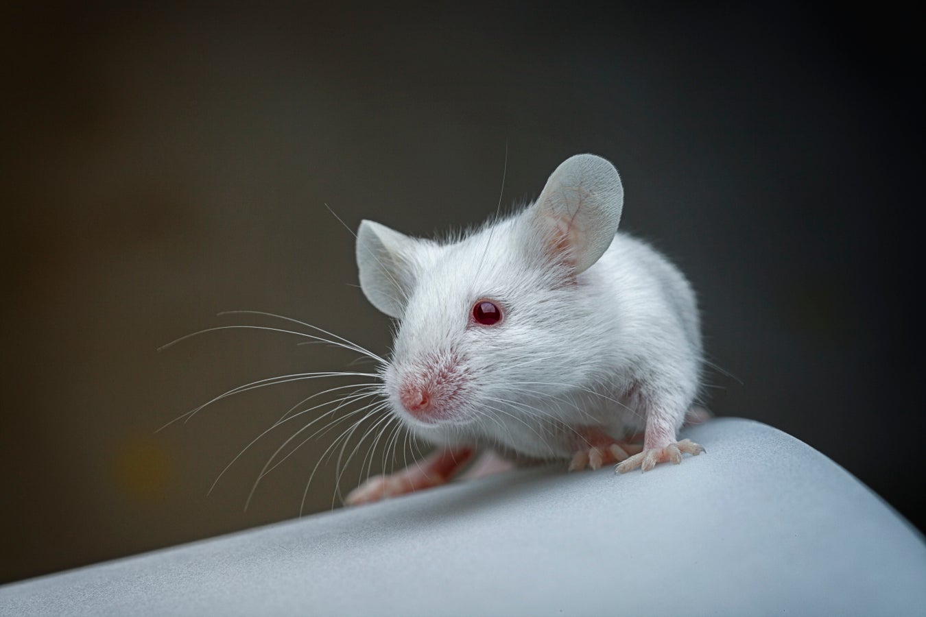
Several research teams have created an atlas of the mouse brain. The map, which has more than 5,300 cell clusters, should help to improve the treatment of brain diseases
By Karin Schlott How many different cell clusters are in the brain of a mouse? Where are they located, and what are their functions? A large international team of researchers has tackled this enormously difficult puzzle and presented a complete cell atlas of the mouse brain.
Experts at the Brain Research through Advancing Innovative Neurotechnologies (BRAIN) Initiative Cell Census Network) documented more than 5,300 different cell clusters, as they have reported in a total of 10 papers published in Nature . As it turns out, cell clusters are distributed in specific regions of the brain and differ depending on their location. This complex structure of the mammalian brain is apparently the result of a long evolutionary process. Hongkui Zeng , director of the Allen Institute for Brain Science in Seattle and a co-author of several of the studies, is convinced that the new brain map will allow researchers to finally see how the brain is organized.
For the scientists, the choice of the brain of the house mouse ( Mus musculus ) is obvious: The rodent is the model organism par excellence in biology and medicine. It furnishes the best way to create an exemplary atlas of a mammalian brain.
The researchers employed various analytic methods for their studies, including different single-cell analyses. These can be used to investigate the genetic activity of brain cells. Among other things, the aim was to find out which messenger RNAs (mRNAs) the mouse brain cells produced. Based on the various RNA snippets, the scientists were then able to deduce how many different cell clusters there are in that brain because each of them produces a characteristic signature of mRNA molecules.
At the same time, the team also determined the spatial position of the cells in the brain. This is what makes this mammoth project so special: all the researchers involved not only mapped “the entire mouse brain” for the first time but also did so using spatial transcriptomics, allowing “cell types to be placed in their native tissue context,” wrote neurologist Maria Antonietta Tosches of Columbia University in an accompanying commentary in Nature .
The researchers sequenced several million cells and collected their location data in the rodent brain. The team then divided the brain cells into four levels comprising a total of 5,322 clusters. The experts also mapped how the individual clusters interact with one another and are in contact with the help of neurotransmitters and neuropeptides.
The 10 studies produced a plethora of results. Deep within the brain—in the areas of the hypothalamus, midbrain and hindbrain—the range of cell types is much greater than in the evolutionarily younger cerebral cortex. “These findings indicate that different rules govern neuronal diversity across the brain, perhaps because each region evolved under different constraints,” Tosches explained in the Nature commentary. The difference could be linked to the fact that the parts of the brain below the cortex have changed less over the course of evolution than the upper parts—possibly because the lower parts control more of the body’s basic functions.
The results of the new studies should also support existing plans to map the human brain . One aim of all this research is to improve the treatment of neurodegenerative diseases and neurological disorders. It is known, for example, that many diseases develop in certain regions of the brain—possibly because specific cell types have changed. According to Zeng, a brain atlas could be used to develop gene therapies or drugs to directly target cells and thus reduce medications’ side effects.
The researchers also hope that their atlas will stimulate numerous other research projects. And finding out the function of the cells and their role in disease may keep the team busy for the next 20 years.
This article originally appeared in Spektrum der Wissenschaft and was reproduced with permission.
Exercise can boost brain health
A fascinating link between regular exercise and better brain health has been revealed, according to an international study that included a team of clinical researchers from Pacific Neuroscience Institute’s Brain Health Center, located at Providence Saint John’s Health Center.
The research, detailed in the paper “Exercise-Related Physical Activity Relates to Brain Volumes in 10,125 Individuals,” was published this week in the Journal of Alzheimer’s Disease and shows being physically active is related to increased size of brain areas important for memory and learning.
The study looked at MRI brain scans from 10,125 people done at Prenuvo imaging centers, a key collaborator in the research.
It found those who regularly engaged in physical activities such as walking, running or sports had larger brain volumes in key areas.
This includes the gray matter, which helps with processing information, and the white matter, which connects different brain regions, as well as the hippocampus, important for memory.
Cyrus A. Raji, M.D., the lead researcher, explains the findings in simple terms: “Our research supports earlier studies that show being physically active is good for your brain. Exercise not only lowers the risk of dementia but also helps in maintaining brain size, which is crucial as we age.”
David Merrill, M.D., study co-author and director of the PBHC noted, “We found that even moderate levels of physical activity, such as taking fewer than 4,000 steps a day, can have a positive effect on brain health. This is much less than the often-suggested 10,000 steps, making it a more achievable goal for many people.”
Study co-authorSomayeh Meysami, M.D. , assistant professor of neurosciences at Saint John’s Cancer Institute and the Pacific Brain Health Center noted, “Our research links regular physical activity to larger brain volumes, suggesting neuroprotective benefits. This large sample study furthers our understanding of lifestyle factors in brain health and dementia prevention . “
A Lancet Study in 2020 found about a dozen modifiable risk factors increase risk for Alzheimer’s disease, including physical activity.
This work builds upon previous work by this group, linking caloric burn from leisure activities to improved brain structure.
“This study demonstrates the influence of exercise on brain health imaging and when added to other studies on the role of diet, stress reduction and social connection offer the proven benefits of drug-free modifiable factors in substantially reducing Alzheimer’s disease,” said George Perry, Editor-in-Chief of Journal of Alzheimer’s Disease.
“With comprehensive imaging scans, our study underscores the interconnected synergy between the body and the brain. It echoes the knowledge of past generations, showcasing that increased physical activity is a predictor of a healthier aging brain,” said Dr. Attariwala, senior author of this paper.
This research highlights an easy way to keep our brains healthy: stay active! Whether it’s a daily walk or a favorite sport, regular physical activity can have lasting benefits for our brain health.
7 Smart Strategies to Boost Your Brain Health in the New Year, According to a Cognitive Neuroscientist
Write these down. Hiroshi Watanabe/Getty Images Looking for actionable ways to stay sharp, maximize your natural brain power, improve mental health, and prevent cognitive decline? Of course you are. The good habits and healthy mindsets we adopt right now really can help protect and maintain our brain health in the long term. But what exactly are the key habits and lifestyle factors that keep the brain “young” and thriving for as long as possible? The answer isn’t as simple as downloading another brain game app ( the science is pretty fuzzy on whether they actually do anything). Instead, cognitive neuroscientist Julie Fratantoni, PhD, head of research and strategic partnerships at Center for BrainHealth , shares the best, science-backed strategies to hack your brain health—and put the brain power you already have to even better use.
Related: 6 Everyday Habits to Train Your Brain to Be Happier Create High-Level Goals—and Set Mini-Goals Along the Way
Setting and working toward a valued goal supports both your sense of well-being and important brain systems. “Having clearly defined goals can strengthen your sense of purpose,” Fratantoni says.
According to Fratantoni, goal-setting is a fantastic exercise for the brain’s frontal network, the region involved in high-level executive functions, reasoning and decision-making, information retrieval, and emotion regulation (just to name a few). “When you engage in setting goals, planning, and prioritization, you’re exercising your frontal network,” she says. “Your frontal lobe is the first part of the brain that’s vulnerable to decline over time, so anything you can do to strengthen it is good.”
Plus, as you take incremental strides toward a larger resolution and accomplish small steps along the way, “you’re activating the brain’s reward network to produce dopamine (which is both motivating and rewarding),” Fratantoni says.
Do focused work toward your goal every day.
Do something that propels you toward the main goal every day for a realistic chunk of time. Fratantoni calls this your “elephant.” “The elephant is not the goal, [but it’s] something you can work on for 45 minutes of focused work that moves you forward,” she says.
Leverage your personal, prime-time mental energy.
Take stock of when you have the most energy and brain power—and then use it to get real stuff done. “This is about being strategic with your mental energy,” Fratantoni explains. “Many people feel sharpest in the morning [and] use that time to answer emails and do other busywork. Instead, block that time to work on a task that will move a major project forward. Save emails and busywork for when you have less energy.”
Related: How to Make Good Habits Stick—Go Beyond New Year’s Resolutions Build Confidence Through Patterns of Positive Self-Talk
It’s time to break free from self-deprecating thought patterns, which trap us in loops of anxiety that limit our cognitive capabilities. Building confidence and generating a positive self-perception (skills that can be learned with practice!) doesn’t just create a more pleasant headspace—it can help change the way your brain works and set you up for better problem-solving, resilience, and innovation.
“When you’re feeling anxious, the anterior cingulate cortex (ACC) narrows your options to fight/flight/freeze, so you are able to act quickly [for survival],” Fratantoni says. “On the other hand, confidence quiets the anxious systems, which allows you to think creatively and solve problems.”
Actively replace pessimistic thoughts with confidence-boosting thoughts.
Instead of fighting to stop negative self-talk (which can seem to have a mind of its own), work on replacing it with confidence-building, self-affirming thoughts . “Telling yourself simple phrases such as ‘I can do this,’ and ‘this can happen,’ help to cultivate a confident mindset,” she says.
Keep some deep breathing exercises in your back pocket.
When your negative thoughts or self-doubt go haywire, Fratantoni recommends practicing a simple slow breathing exercise (inhale for four counts, exhale for six counts) to get your system out of anxiety mode and “bring your rational brain back online.”
More easy breathing exercises to try: Box Breathing
4-7-8 Breathing
Mindful Breathing Series
Related: Want to Be More Positive? Here Are 7 Things Optimistic People Do Differently Get Your Heart Rate Up Routinely
It really can’t be overstated how beneficial exercise is for brain health and mood management —not just your heart, bones, joints, and muscles. Prioritize getting a daily dose of heart-pumping movement to increase blood flow to the brain and boost cognition .
“Moving your body and raising your heart rate improves thinking, learning, problem-solving and emotional balance,” Fratantoni says. “Physical exercise sparks the birth of new neurons in the hippocampus, which is the part of the brain that is responsible for memory and learning . [It also] increases brain derived neurotropic factor (BDNF) , which sparks the birth of new neurons.”
Find ways to move that don’t intimidate you—and that you’re likely to return to consistently.
Just a small increase in physical activity (even if you’re starting at zero!) is all it takes to see some benefits. So, think small. Walking is a fantastic form of exercise , especially if you can pick up the pace a little bit, or walk on an incline , to the point where you’re breathing a bit heavier and raising your heart rate. Even vigorous gardening or household chores , the kind of tasks that make you want to strip down to just a T-shirt—like vacuuming or cleaning out the garage—all count.
Advertisement
“Small changes can make a big impact,” Fratantoni says. “Taking the stairs, going on a 10-15 minute walk every day , and even playing with your kids are all small but important ways to keep you active throughout the day that don’t require spending hours in the gym.” Boost Your Memory: Challenge Yourself to Think Even Deeper Memory decline is a normal part of getting older, but it doesn’t always need to happen so soon or so rapidly. Wondering how to “workout” your memory “muscles” for better information retention and recall? Challenge yourself to expand upon the info you’ve taken in.”After you engage with information (like a podcast, article, or […]
Guayusa tea and lion’s mane supplements could improve cognitive function

Supplements derived from the fungus known as as lion’s mane (pictured above) and guayusa tea could help improve mood and cognitive function. Image credit: Alex Ratson/Getty Images. Guayusa and lion’s mane are two natural dietary supplements that may have health benefits.
Usually taken as a tea made from the leaves, guayusa is a plant in the Ilex (holly) family. Lion’s mane is a mushroom, used for culinary purposes, which can also be taken as a supplement.
Studies have suggested that both have antioxidant and anti-inflammatory properties, alongside other health benefits.
A new study has found that they might also enhance mood and cognitive function.
Many people take supplements to enhance their health and wellbeing, but while animal studies have demonstrated their positive effects, there is little conclusive evidence of their efficacy in people.
Guayusa is from a tree in the holly family, Ilex guayusa , which grows widely in South America. People have been drinking it as a tea in the western Amazon for centuries. The plant contains many polyphenols and alkaloids , one of which is caffeine.
Potential health benefits of guayusa include: antioxidant activity
anti-inflammatory effects
cardiovascular protection
anti-obesity and anti-diabetic effects
neuroprotective activity.
Lion’s mane is a white, spherical mushroom ( Hericium erinaceus ). In Asia, it is used for both culinary and medicinal purposes, and it is becoming increasingly available as a supplement, because of its reported health benefits. The extract can be taken as capsules, liquid, tablet or powder.
Studies have suggested that benefits of lion’s mane may include: antioxidant and anti-inflammatory properties
supporting immune function
relieving anxiety and depression
lowering cholesterol
cancer prevention or treatment
controlling blood sugar
cognitive benefits.
Until now, evidence for these benefits has been largely from animal studies.
A new study now suggests that both supplements may enhance mood and cognition in people. The study is published in the journal Nutrients .
Kelsey Costa , a registered dietitian and nutrition consultant for the National Coalition on Healthcare, not involved in this research, commented on the study for Medical News Today : “The study’s findings align with previous research suggesting cognitive benefits from these natural compounds. However, we should approach these results with cautious optimism given the study’s limited size and funding source — Applied Food Sciences Inc., the makers of AMT [the guayusa supplement]. This introduces the potential for bias in the study design and interpretation of results.” Small study looks at herbal supplements for brain health
A total of 40 people took part in the study — 22 female and 18 male particiapants. All were aged between 18 and 50 years, were in good health, had a body mass index (BMI) between 18.5–39.9 kilograms per square meter (kg/m 2 ), and were habitual consumers of up to 240 milligrams (mg) of caffeine — equivalent to around 2–3 cups of coffee — per day.
Participants underwent one screening visit and three test visits. At the screening visit, researchers assessed their health and diet, and participants did practice neuropsychological assessments to familiarize themselves with the processes for the test visits.
For 24 hours before each test visit, participants had to eat exactly the same diet and avoid exercise and alcohol. In addition, they were told to avoid caffeine for 12 hours, and fast for 8 hours before the test visit.
On arrival for each test visit, they were randomly allocated to receive placebo, 650 mg guayusa extract (AMT: AmaTea® Max), or 1 gram (g) Nordic-grown lion’s mane.They completed baseline testing to assess mood, focus, mental clarity, concentration, ability to be productive and tolerate stress, and happiness (using the Subjective Happiness Scale ), before taking the supplement, after 60 minutes and after 120 minutes.At each time point, participants also underwent neuropsychological assessments, which assessed reaction time, mental processing, cognitive control, and attention. Researchers also checked their blood pressure and heart rate. Guayusa and lion’s mane improved mood and cognition Researchers found that both supplements improved reaction time, with lion’s mane having a greater effect at 120 minutes than 60, suggesting a delayed response after ingestion.The two supplements had some different effects, with lion’s mane appearing to have some more lasting effects than guayusa. This finding is backed up by a study that suggests lion’s mane given over an 8-week period decreased depression, anxiety, and sleep disorders in a group of people with overweight or obesity. Guayusa significantly improved mental clarity, focus, concentration, mood, and productivity at 60 and 120 minutes post ingestion, and the ability to tolerate stress at 60 minutes. Lion’s mane improved subjective ratings of happiness at 60 minutes and this effect persisted to 120 minutes, whereas guayasa improved this only at 120 minutes. The researchers caution that guayusa did raise both diastolic and systolic blood pressure by, on average 5mmHg, but that it remained within the normal range for healthy blood pressure. Learn About Healthy Eating Explore Our Science-Backed Nutrition Resources How might guayusa and lion’s mane have these effects? “Guayusa has caffeine and similar derivatives. These are essentially stimulants, which we know can boost cognitive performance,“ Prof. Maryann Amirshahi , medical director of the National Capital Poison Center, not involved in this study, explained to MNT . “Guayusa and lion’s mane also contain antioxidants — these are chemicals that prevent damage to nerves and other cells. There are many different chemicals in both of these, so it is hard to say which one, or a combination of them is what boosts cognitive performance,” she noted. Prof. Amirshahi nevertheless cautioned that the study had some limitations: “An important limitation of a recent study is that they only looked at cognitive performance at 2 hours. We do not know how long these effects last and if they are offset by poor sleep. We also do not know how effective or safe they are with longer term use.”Costa further explained some of the effects: “As noted by the authors, caffeine appears as a key factor in the observed cognitive improvements associated with AMT. […] Essentially, caffeine boosts alertness […]
New study shows exercise can boost brain health

Credit: CC0 Public Domain A fascinating link between regular exercise and better brain health has been revealed, according to an international study that included a team of clinical researchers from Pacific Neuroscience Institute’s Brain Health Center, located at Providence Saint John’s Health Center.
The research, detailed in the paper “Exercise-Related Physical Activity Relates to Brain Volumes in 10,125 Individuals,” was published in the Journal of Alzheimer’s Disease and shows being physically active is related to increased size of brain areas important for memory and learning.
The study looked at MRI brain scans from 10,125 people done at Prenuvo imaging centers, a key collaborator in the research. It found those who regularly engaged in physical activities such as walking, running or sports had larger brain volumes in key areas. This includes the gray matter , which helps with processing information, and the white matter , which connects different brain regions, as well as the hippocampus, important for memory.
Cyrus A. Raji, M.D., the lead researcher, explained, “Our research supports earlier studies that show being physically active is good for your brain. Exercise not only lowers the risk of dementia but also helps in maintaining brain size , which is crucial as we age.”
David Merrill, M.D., study co-author and director of the PBHC noted, “We found that even moderate levels of physical activity , such as taking fewer than 4,000 steps a day, can have a positive effect on brain health. This is much less than the often-suggested 10,000 steps, making it a more achievable goal for many people.”
Study co-author Somayeh Meysami, M.D., assistant professor of neurosciences at Saint John’s Cancer Institute and the Pacific Brain Health Center noted, “Our research links regular physical activity to larger brain volumes, suggesting neuroprotective benefits. This large sample study furthers our understanding of lifestyle factors in brain health and dementia prevention.”
A Lancet Study in 2020 found about a dozen modifiable risk factors increase risk for Alzheimer’s disease, including physical activity. This work builds upon previous work by this group, linking caloric burn from leisure activities to improved brain structure.
“This study demonstrates the influence of exercise on brain health imaging and when added to other studies on the role of diet, stress reduction and social connection offer the proven benefits of drug-free modifiable factors in substantially reducing Alzheimer’s disease,” said George Perry, Editor-in-Chief of Journal of Alzheimer’s Disease.
“With comprehensive imaging scans, our study underscores the interconnected synergy between the body and the brain. It echoes the knowledge of past generations, showcasing that increased physical activity is a predictor of a healthier aging brain,” said Dr. Attariwala, senior author of this paper.
This research highlights an easy way to keep our brains healthy: stay active! Whether it’s a daily walk or a favorite sport, regular physical activity can have lasting benefits for our brain health.
Provided by IOS Press
There’s a ‘Wave of Death’ in Every Human Brain. Scientists Are Scrambling to Stop It.

Working to Stave Off Brain’s ‘Wave of Death’Ioannis Tsotras – Getty Images Researchers studying the brain’s final moments have gained new insight into the “wave of death” that occurs before a brain’s activity fully flatlines.
When neural activity stops, it doesn’t stop abruptly, but over time.
The team hopes to find ways to keep the brain functioning even when the heart and lungs fail.
The brain doesn’t shut off like a light switch, even as death approaches. While other bodily organs —namely the heart and lungs—have sudden stops, the brain flickers on through active neurons in a “wave of death ” until it reaches a state of electrical silence.
Brain researchers in Paris have been working to better understand these cascading changes that occur when a brain is deprived of oxygen , as well as what they mean for our conceptualization of death. The results, published in the journal Neurobiology of Disease , show where this wave originates and just how we can potentially stave it off.
Stephane Charpier, head of the research team at the Paris Brain Institute and study author, said in a statement that her team now knows “that a flat EEG does not necessarily mean the definitive cessation of brain functions.” This means that there could be hope for our ability to keep an oxygen-starved brain working even longer than expected.
When the brain stops receiving oxygen, the adenosine triphosphate (ATP) stores that act as fuel for the cells quickly get sucked away. Then, it all becomes a bit chaotic. Electrical balances go out of whack and huge amounts of chemicals are released. “Neural circuits seem to shut down at first,” Severine Mahon, another author on the paper, said in a statement, “then we see a surge in brain activity—specifically an increase in gamma and beta waves. These waves are usually associated with a conscious experience. In this context, they may be involved in near-death experiences reported by people who have survived cardiorespiratory arrest.”
Once the process starts, the neuron activity trends downward until the brain is completely electrically silent. But that isn’t the end, because it’s now time for the “wave of death,” which totally alters the function and structure of the brain. “This critical event, called anoxic depolarization, induces neuronal death throughout the cortex,” Antoine Carton-Leclercq, first author on the study, said in a statement. “Like a swan song, it is the true marker of transition toward the cessation of all brain activity.”
Advertisement
And now we know where it’s coming from. The team found (while studying rats ) that the wave originates in a part of the brain called the neocortex—a region that makes up a large percentage of your brain, which can be divided into six layers. The “wave of death” seems to originate in layer five, deep in the tissue of the neocortex. “We have observed this same dynamic under different experimental conditions,” Mahon said, “and believe it could exist in humans.”
Why does this phenomenon come from so deep in the brain? The team believes that this “wave of death” starts in layer five because the neurons in that layer have exceptionally high oxygen requirements. But it seems like restoring oxygen flow can reverse at least some of these effects. When the team reoxygenated the rats’ brains, the cells replenished their ATP reserves and restored synaptic activity.
And that means there’s hope that the wave can be stopped . Carton-Leclercq said that researchers already knew that brain function could be protected if a patient was resuscitated fast enough. But—while it may take time to figure out exactly how to stop this wave—knowing where it comes from could eventually be helpful in preserving function even more effectively.
“This new study advances our understanding of the neural mechanisms underlying changes in brain activity as death approaches,” Charpier said in a statement. “It is now established that, from a physiological point of view, death is a process that takes its time… and that it is currently impossible to dissociate it rigorously from life.”
“We now need to establish the exact conditions under which these functions can be restored,” Charpier continues, “and develop neuroprotective drugs to support resuscitation in the event of heart and lung failure.”
You Might Also Like
Study links tiny pieces of plastic in the environment to Parkinson’s and Alzheimer’s diseases

It is no secret that the plastics in our environment are making us ill, but recent research demonstrates the power of a specific type of plastic to destroy our brains and cause diseases such as Parkinson’s and Lewy body dementia. The study was carried out by scientists from Duke University and published in the journal Science Advances .
The plastics in question are nanoplastics, which are miniscule particles of plastic that come from common plastic objects that we use in our everyday lives. In particular, the researchers found that polystyrene nanoparticles, which come from plastic utensils, plastic cups and similar items, bind to a protein known as alpha-synuclein which has been linked to Lewy body dementia and Parkinson’s disease.
Scientists have observed buildups of aberrant types of this protein in Parkinson’s patients. Alpha-synuclein is believed to serve an important function in keeping neurons in the brain healthy , but when it becomes misfolded or warped, it can lead to problems. Scientists do not fully understand yet whether this is a cause or a symptom of Parkinson’s.
The researchers witnessed this type of accumulation in cultured neurons, test tubes and mouse models. The study’s lead author, Andrew West, said that the team’s most surprising discovery while conducting their research was the strong connections formed between the protein and plastic within neuron lysosomes. These are digestive organelles that allow cells to break down waste products with support from enzymes. Evidence shows that plastic therefore interferes with neurons’ natural cleaning processes.
West noted in a statement: “Our study suggests that the emergence of micro and nanoplastics in the environment might represent a new toxin challenge with respect to Parkinson’s disease risk and progression.”
Although further studies are needed to explore this connection further, this study is just the latest in a large body of epidemiological and lab research demonstrating how the environment is playing a role in the rise in Parkinson’s disease being seen right now.
Parkinson’s is currently the fastest-growing neurological disorder worldwide. Unfortunately, the presence of these plastic toxins in our water and food supply is only expected to grow, which means that many more people could be at risk of developing this devastating illness in the near future.
Scientists do not fully understand what causes Parkinson’s, although they have plenty of evidence that exposure to certain pollutants and toxins are risk factors for the disease. While many studies have focused on pesticides and chemicals, few have looked into the role nanoplastics could play in the development of the disease. Plastics in the environment must be studied in depth to determine their role in illness
There is a growing body of evidence that nanoplastics can be found throughout our environment, especially in indoor air. When people inhale them, they enter the brain and the bloodstream directly, raising people’s risk of cancer.
Some experts have pointed out that previous environments have a big influence on people’s current health. Just like previous smoking habits raise a person’s risk of lung cancer, living environments replete with nanoplastics can raise a person’s risk of cancer, Parkinson’s and Alzheimer’s.
It is an unfortunate reality that most people are already carrying microscopic plastic particles in their blood, and determining how that is affecting our health will be a key area of scientific research in the coming years.
West stated: “While microplastic and nanoplastic contaminants are being closely evaluated for their potential impact in cancer and autoimmune diseases, the striking nature of the interactions we could observe in our models suggest a need for evaluating increasing nanoplastic contaminants on Parkinson’s disease and dementia risk and progression.”
Sources for this article include:
GreatGameIndia.com
ScienceAlert.com
Brainoware: A breakthrough AI approach using brain organoids for advanced computation
In a study published in the journal Nature Electronics , researchers in the United States of America developed an artificial intelligence (AI) hardware approach named “Brainoware” that employs adaptive reservoir computation of the brain organoid neural networks (ONNs). They found that the approach could achieve nonlinear dynamics, fading memory properties, and unsupervised learning, demonstrating its practical potential in tasks such as nonlinear equation prediction and speech recognition.
Study: Brain organoid reservoir computing for artificial intelligence. Image Credit: Dragon Claws / Shutterstock Study: Brain organoid reservoir computing for artificial intelligence . Image Credit: Dragon Claws / Shutterstock Background
Silicon computer chips majorly drive the artificial neural networks (ANNs) that form the basis of AI. However, training ANNs on these chips has limitations such as high energy and time consumption, the Neumann bottleneck (physical segregation of data from data processing), and slowing of Moore’s law (slower doubling of transistors in an integrated circuit). This emphasizes the need to design new AI hardware approaches.
The human brain, with its complex three-dimensional network of cells and synapses, inspires the development of AI hardware due to its energy efficiency, neurogenesis, and neuronal plasticity that enable the processing of noisy data at minimal training cost. While neuromorphic chips attempt to mimic brain functions, there is a need to enhance their capabilities in terms of processing information, addressing real-life uncertainty, and utilizing energy.
Brain organoids are three-dimensional aggregates developed in vitro using stem cells to mimic brain structure and function. Researchers in the present study developed a novel AI hardware system named “Brainoware” using brain organoids, which harnesses reservoir computation and learning abilities of neural networks of the organoid. About the study
Brainoware was integrated into a reservoir computing framework with three main components: an input layer, a reservoir layer (organoid), and an output layer. The system was constructed by mounting a functional brain organoid, developed using human pluripotent stem cells, onto a high-density multielectrode array (MEA). The organoid formed the reservoir layer and showed the presence of various brain cell types, early brain-like structures, and network electrical activity. The organoid received signals via the input layer, which converted time-dependent inputs into spatiotemporal sequences of electric stimulation. The brain organoid acted as an “adaptive living reservoir” that mapped these signals to the ONNs. In the output layer, the MEA recorded neural activities representing the reservoir state (using techniques like linear or logistic regression), and decoded them to provide readouts for applications like classification, recognition, and prediction.
The physical reservoir properties of Brainoware were tested, including nonlinear dynamics, spatial information processing, and fading memory, by assessing the ONN’s response to stimulations of varying pulse times and voltages. The system was then applied to real-world tasks such as speech recognition and nonlinear chaotic equation prediction.
In the speech recognition task, Brainoware was required to identify the speaker’s vowels in a speaker pool. A total of 240 audio clips of isolated Japanese vowels pronounced by eight different male speakers were used to train the system, and the evoked responses were classified by logistic regression. Functional connectivity changes in the organoid before and during training were measured separately. In the next task, Brainoware was made to predict the Hénon map, a nonlinear dynamic system with chaotic behavior. A two-dimensional Hénon map was decomposed to one dimension and used as an input to Brainoware. The evoked activity was recorded before and during training and compared with controls. Results and discussion
Brainoware showed the properties of a physical reservoir. The nonlinear response, spatial processing, and fading dynamics of ONNs could be controlled by modifying the input stimulation parameters. Stronger responses and slower relaxation were observed with high voltage pulses and long duration. The dynamics of Brainoware were found to be in accordance with that of ANNs and memristors. Through training, Brainoware was found to enhance its computational performance and exhibit unsupervised learning capabilities through its adaptive living reservoir.
Brainoware improved its accuracy over training from 51% to 78% in the speech recognition task, suggesting the triggering of unsupervised learning abilities of the organoid via electrical stimulation. Test results indicated that training reshaped the functional connectivity of the organoid, thus initiating unsupervised learning.
Additionally, Brainoware could successfully predict the Hénon map and outperformed other methods, such as linear regression and ANN, with a long short-term memory unit. Brainoware could not perform without the organoid, as indicated by the nil regression score of the organoid-less control system. The performance was found to improve with training, as the improved regression scores indicated. Test results showed that Brainoware’s learning activity was dependent on neural plasticity. Conclusion
In the present study, researchers introduced Brainoware as a reservoir computing hardware that harnesses the computational power of brain organoids and demonstrated its adaptability, plasticity, and potential to address challenges in current AI hardware. Despite being highly promising, the Brainoware approach encounters challenges in organoid generation and maintenance, power consumption by the peripheral equipment, the use of flat and rigid MEAs, and a lack of efficient data management tools. In the future, customized, low-power, brain-inspired systems could be developed with advanced brain-machine interfaces and data management software for improved applicability and accuracy.
80 Percent of Store Receipts Contain Bisphenol – Test Results: Receipts From 2022
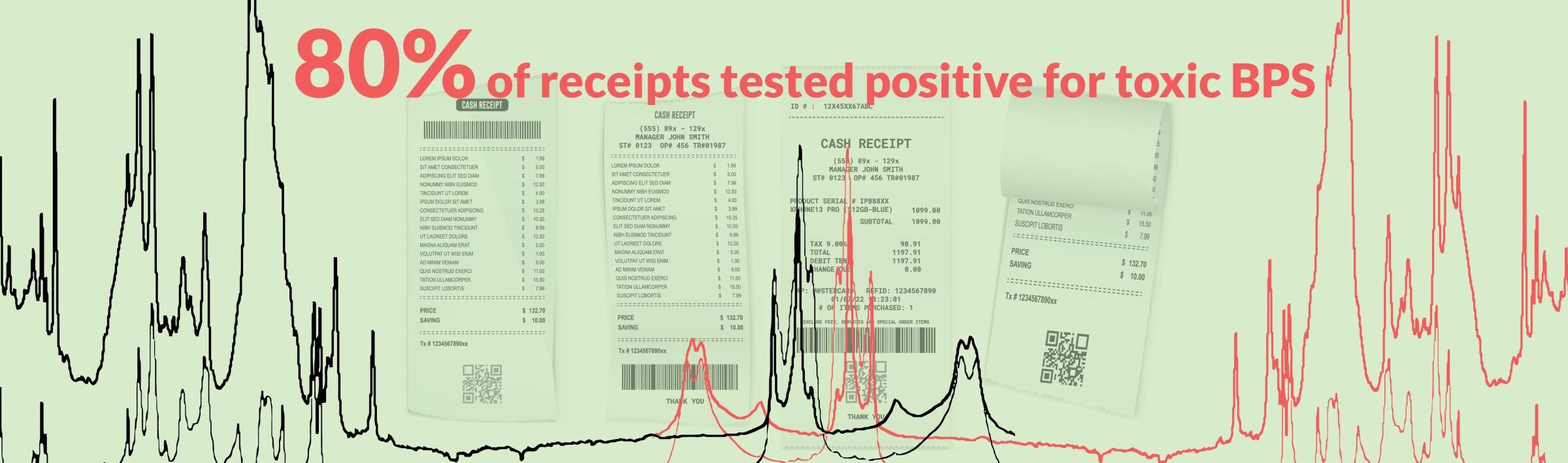
The table shows test results from purchase receipts printed at national and large regional chain stores in the United States in 2022.
BPS = Bisphenol S
BPA = Bisphenol A
Business Receipts Tested Store Locations Results 7-Eleven 1 MI BPS Ace Hardware 4 MI BPS ALDI 14 MD, MI, MN, NC, NJ BPS Altar’d State 1 MN BPS Amazon 1 Unknown BPS American Eagle 1 CA BPS Apple Store 1 MI BPS Athleta 2 IL, MI BPS Auto Zone 2 MD, MN BPS Bahama Breeze 2 DE BPS Barnes & Noble 1 MI BPS Baskin Robbins 1 MI BPS Bed Bath and Beyond 2 MI, VA BPS Best Buy 5 MD, MI, MN, VA Non-bisphenol Big Lots 1 MD BPS Bojangles 1 TN BPS Boston Market 1 NJ BPS BP Sunrise 1 FL BPS Burger King 1 MI BPS Carter’s 1 MI BPS Casey’s General Store 1 MN BPS Cenex Travel Plaza 2 MN BPS Chick-fil-A 1 MD, TN BPS Chipotle 5 CA, D.C., IL, MI BPS Circle K 4 MI BPS Clothes Mentor 1 MI BPS Costco 1 MN BPS Costco 5 MI, MN, WA Non-bisphenol Costco Gas 1 MI BPS Culver’s 1 MN Uncoated CVS 1 MN BPS CVS 7 MA, MI, MN Non-bisphenol Dairy Queen 1 MN BPS Dick’s Sporting Goods 1 MI BPS Dollar General 1 MI BPS Dollar Tree 6 MI, MN, NJ BPS Domino’s Pizza 1 MN BPS DSW 1 MN BPS Dunham’s Sports 1 MI BPS Dunkin Donuts 1 MI BPS Exxon Cross Roads Gas and C-store 2 MD BPS Family Dollar 3 MD, MI BPS FedEx 1 MI BPS Five Below 3 MI, NJ BPS Food Lion 3 GA, NC, VA BPS Forever 21 1 MI BPS FP Movement (Free People) 1 CA BPS Fresh Thyme 2 MI BPS Gap Outlet 1 MD BPS Giant 3 MD, VA BPS H Mart 1 MA BPS H&M 1 MN Non-bisphenol Hannaford 1 ME BPS Harbor Freight 1 MI BPS Harris Teeter 2 D.C., NC BPS Holiday Station Store 3 MN BPS Home Goods 3 MI, NJ Non-bisphenol Hot Topic 1 MI BPS Huntington Bank 1 MI BPS Hy-Vee 1 MN BPS IKEA 1 MI BPS Illy Coffee 1 MI BPS In-N-Out Burger 1 CA BPS Jamba Juice 1 MN BPS JCPenney 2 MD BPS Jersey Mike’s Subs 1 MD BPS JoAnn Fabric and Crafts 3 MI, MN BPS KFC 1 MN BPS Kohl’s 1 MI BPS Kroger 19 MI BPS Kroger Fuel 1 MI BPS Kwik Trip 2 MN BPS L.L. Bean 1 MN BPS Lego 1 NY BPS Little Caesar’s 1 MI BPS Lowe’s 5 MD, MI, NJ BPS Lululemon 1 CA Non-bisphenol Macy’s 1 MI BPS Marshalls 1 MI Non-bisphenol McDonald’s 7 MI, MN, TN, VA BPS Meijer 7 MI BPS Meijer Gas Station 2 MI BPS Menards 5 MI, MN BPS Michaels Craft Store 2 MN, NJ BPS NAPA Auto Parts 1 MN BPS Noodles & Company 2 MI BPS Nordstrom 1 MI BPS Nordstrom Rack 1 MI BPS Ocean State Job Lot 2 MA BPS Office Depot 2 MD, MI BPS Old Navy 2 CA, MD BPS Olive Garden 2 NJ BPS One Stop 2 WV BPS Panda Express 7 MD BPS Panda Express 2 MD Inconclusive Panera Bread 2 MI BPS Party City 1 MI BPS Petco 2 CA, MN BPS PetPeople 1 MI BPS PetSmart 3 MI, MN, NJ BPS PGA Tour Superstore 1 MN BPS Pho Hoa 1 MA BPA Pilot 2 KY, SC BPS Plum Market 2 MI BPS PNC Bank 1 MI BPS Popeye’s 6 MD BPS Ragstock 2 MI, MN BPS Ralphs 1 CA BPS REI 2 MI, WA Non-bisphenol Rite Aid 6 MI, NY BPS Runnings 2 MN BPS Sally Beauty 1 MD BPS Sam’s Club 3 MN, NV BPS Sam’s Club Gas 2 NV Non-bisphenol Sephora at JC Penney 1 MI BPS Shake Shack 1 MI BPS Shell 4 MD, MI BPS Sherwin-Williams 2 MI BPS ShopRite 2 NJ BPS Sierra Trading Post 1 MI Non-bisphenol Smith’s Food and Drug 1 NV BPS Speedway 1 MN BPS Spencer’s 1 MI BPS Staples 1 MI BPS Starbucks 2 MI, WA Non-bisphenol Subway 1 MI BPS Taco Bell 2 MN, WV BPS Target 17 MI, MN, NJ, NY, VA, WA Non-bisphenol The Home Depot 8 MA, MD, MI, MN, NJ, VA BPS The UPS Store 1 MN BPS The Vitamin Shoppe 1 MI BPS Tim Horton’s 1 MI BPS TJ Maxx 1 MI Non-bisphenol Trader Joe’s 13 CA, MA, MD, MI, MN, NC, NV Non-bisphenol Ulta 1 MI BPS United States Postal Service 12 MI, MN BPS Universal Studios 2 FL BPS Verizon 1 NY BPS Victoria’s Secret 1 MI BPS Walgreens** 6 MI, MN, NY, WA BPS Walmart 11 FL, GA, MA, MD, MI, MN, NJ BPS Wegmans 5 MA ,NJ, NY BPS Wendy’s 3 MN BPS Whole Foods Market 3 CA BPS Whole Foods Market 13 CA, FL, MA, MI, WA Non-bisphenol WinCo Foods 1 NV BPS Wyndham Garden Hotel 1 MI BPS
Study: Citrus fruits can help you maintain healthy cognitive function as you age

Advertisement
Organic fruits are rich sources of essential vitamins and minerals and beneficial plant compounds. They are also loaded with dietary fiber, which you need to maintain a healthy heart and digestive system, as well as healthy blood sugar levels. Because they’re packed with all these health-giving components, it’s not surprising that experts recommend eating a variety of organic fruits as part of a healthy, well-balanced diet.
Older adults, in particular, can get considerable benefits from eating fruits every day. Decades of research show that consuming citrus fruits like lemon, orange, grapefruit and the like, can help the elderly maintain their cognitive abilities . It is a well-established fact that cognitive functions naturally decline as people age. But scientific evidence suggests that eating citrus fruits could help slow down this natural process and keep older adults mentally sharp. Citrus flavonoids can boost the aging brain
Flavonoids are the main polyphenols in raw citrus fruits. Naturally occurring chemical reactions convert these compounds into various forms, such as flavanones, flavonols, flavones and anthocyanins. In citrus fruits, flavanones are the most predominant flavonoids and are said to be responsible for many of the health benefits these fruits offer. Some examples of citrus flavanones include hesperidin (cardioprotective), narirutin (anti-inflammatory), didymin (anticancer) and eriocitrin (antioxidant).
According to a study published in the journal Frontiers in Neuroscience , flavanones are also behind the wonderful brain benefits of citrus fruits. Researchers have found that these flavanones can cross the blood brain barrier and exert their antioxidant effects directly on the brain, mainly by scavenging free radicals to prevent oxidative stress. Oxidative stress, which can damage or kill brain cells, is heavily implicated in brain aging and neurodegenerative diseases .
Inflammation, which is triggered by oxidative stress, is also linked to age-related cognitive impairment. In fact, the brains of people with Parkinson’s and Alzheimer’s disease show heavy signs of brain inflammation . But preclinical studies show that citrus flavanones can protect against brain inflammation by reducing the production of pro-inflammatory chemicals.
This appears to be one of the ways citrus flavanones help preserve memory function in older adults. As reported by a study published in the journal Neurology , adults in their 40s, 50s and 60s with high levels of inflammatory markers tend to experience a steeper decline in thinking and memory skills in their later years than those with low levels of inflammation. This suggests that inflammation is a huge contributor to the decline in cognitive function experienced by older adults.
In addition to memory, flavanone-rich citrus fruits can also help improve executive function, perceptual speed and visuospatial skills in the elderly. This was reported in the Hordaland Health Study, which included over 2,000 Norwegian adults aged 70 to 74 years. The study found that participants with the highest intakes of plant-based foods, particularly citrus fruits, performed significantly better in cognitive tests than those with the lowest intakes.
In a separate study, researchers from the U.K. investigated the cognitive benefits of drinking orange juice enriched with flavanones like hesperidin and narirutin. They found that middle-aged adults (30 to 65 years) who took the drink experienced improvements in psychomotor speed (i.e., the ability to process new information and respond to it physically) after two hours, and in attention/alertness and executive function after six hours.
Meanwhile, a notable study involving over 13,000 elderly Japanese found that consuming citrus fruits three to four times a week can help lower the risk of dementia by 23 percent . (Related: Regular consumption of tea found to reduce risk of dementia and improve cognitive function .)
Citrus fruits are great sources of plant compounds that can help delay brain aging. They are also rich in vitamin C, a potent antioxidant that can boost immune function and support a healthy brain .
Learn more about citrus fruits and other brain foods at Superfoods.news .
Watch this video to learn about how citrus fruits can help prevent dementia . Eating more citrus fruits is an easy way to prevent dementia
This is a modal window.
No compatible source was found for this media.
This video is from the Natural News channel on Brighteon.com . More related stories:
Citrus fruits can counteract some of the health risks of obesity .
Increasing your daily intake of fruits and vegetables can truly help you live longer, reveals study .
Studies show antioxidant-rich foods can help reduce risk of skin cancer .
Support brain health and improve cognitive function with these herbs and spices .
Study: Drinking oolong tea can help reverse aging .
Sources include:
FrontiersIn.org
Memory.UCSF.edu
ScienceDirect.com
Magazine.HMS.Harvard.edu AARP.org Cambridge.org 1 Link.Springer.com Cambridge.org 2 Brighteon.com
Brainoware: Organoid Neural Networks Inspire Brain-AI Hardware
Credit: BlackJack3D / iStock / Getty Images Plus “The human brain has 100 billion neurons, each neuron connected to 10,000 other neurons. Sitting on your shoulders is the most complicated object in the known universe.” — Michio Kaku, PhD.
Since most examples of brain-inspired silicon chips are based on digital electronic principles, their capacity to fully imitate brain function is limited. Self-organizing brain organoids connected to microelectrode arrays (MEAs) can be changed in function to create neural networks. These networks, called organoid neural networks (ONNs), show the capacity for unsupervised learning, which is what artificial intelligence (AI) is based on. These mini-organs, when connected to the right hardware, can even be trained to recognize speech.
This brain-inspired computing hardware, or “Brainoware,” could overcome existing shortcomings in AI technologies, providing natural solutions to challenges regarding time and energy consumption and heat production of current AI hardware. These ONNs may also have the necessary complexity and diversity to mimic a human brain, which could inspire the development of more sophisticated and human-like AI systems.
Brainoware was developed in the labs of Feng Guo, PhD, at Indiana University Bloomington and Mingxia Gu, MD, PhD, at Cincinnati Children’s Hospital Medical Center. The findings were published in the research article “ Brain organoid reservoir computing for artificial intelligence ” in Nature Electronics . Organoid neural networks
Hongwei Cai, the first author of Brainoware, and his colleagues wanted to find a biological way to solve the problem of reservoir computing. Reservoir computing is known for its unique way of processing and learning complex temporal and sequential data. Reservoir computing enables the extraction of intricate patterns and relationships from temporal sequences. This approach ensures fast training and emphasizes energy efficiency, making it a viable option for environmentally conscious AI solutions.
Reservoir computing has demonstrated promising outcomes across various applications, including time-series prediction, speech recognition, language modeling, and addressing complex nonlinear dynamical systems. Its unique architecture and training methodology offer an innovative alternative for effectively managing sequential and temporal data. This renders it a valuable tool within machine learning and artificial intelligence research domains.
Will the computers of the future be made of brains?
Not quite yet. While Brainoware doesn’t require much power consumption, it depends on things like incubators and some systems, such as skilled cell culture technicians or automated systems, for maintaining the cell culture.
Additionally, the generation of organoids is still a relatively uncontrolled, heterogeneous mix of cell types, both dead and alive. Recent engineering efforts to improve the conditions for organoid differentiation and growth, as well as changing their microenvironments, could make it possible to make and keep a lot of standardized organoids.
Data management and analysis is another technical challenge. Data interpretation, extraction, and processing improvements from multiple sources and modalities are still needed to optimize the encoding and decoding of temporal information to and from Brainoware. The power of Brainoware is all but useless without the development of new algorithms and methods for analyzing and visualizing the data.
Although it may be decades before general biocomputing systems are developed, this research will likely yield fundamental insights into learning mechanisms, neural development, and the cognitive implications of neurodegenerative diseases. It could also aid in developing preclinical models of cognitive impairment for testing new therapeutics.
Can wasabi help your memory? A new study has linked the sushi condiment to a better brain

Nigiri is topped with pickled wasabi, which has long been known as an antioxidant. Most recently it’s been linked with improved memory. Sushi lovers, listen up.
New research has found that wasabi is much more than a condiment with a kick. It’s active ingredient, 6-MSITC , is now not only a known antioxidant and anti-inflammatory, but also has been linked to improved short- and long-term memory.
The study, published recently in the journal Nutrients by researchers at Tohoku University, split 72 healthy volunteers between the ages of 60 and 80 into two groups. One received 100 milligrams of wasabi extract and the other got a placebo pill with zero wasabi.
After three months of daily use, this is what was found: Wasabi’s newly discovered benefit
After a few short months, those given 100 milligrams of wasabi daily were tested on language skills, concentration and ability to carry out simple tasks. All were showing boosts in memory, both short- and long-term.
“We knew from earlier animal studies that wasabi conferred health benefits,” lead researcher Rui Nouchi, an associate professor at Tohoku University’s Institute of Development, Aging and Cancer, told CBS News.
“What really surprised us was the dramatic change. The improvement was really substantial,” Nouchi shared.
Episodic memor y – the ability to learn, store, and retrieve information – increased by an average of 18% in the wasabi subjects, who also scored on average 14% higher overall than those given the placebo pill.
6-MSITC, again, known for its anti-inflammatory benefits , seems to reduce inflammation in the area of the brain responsible for memory – the hippocampus.
What is a healthy diet? Why the ‘healthiest’ one considers things other than food. Wasabi could be breakthrough for dementia patients
Nouchi pursued his research because of his work around dementia prevention, CBS News reported.
The recommended methods to improve brain health, such as certain diets and exercise, seem too involved for seniors to follow, Nouchi believes.
Adding a daily supplement would be easier for at-risk seniors to stick with and offer more benefits than other spices used similarly, such as ginger and turmeric, he believes.
The Tohoku team will be testing wasabi on younger groups in preparation for testing on dementia patients. Pure wasabi is hard to find
CosMc’s: McDonald’s reveals locations for chain’s new spinoff restaurant and menu Krispy Kreme reveals ‘Elf’ collection before ‘Day of the Dozens’ deal: How to get a $1 box Popeyes throws shade at competitors with ‘DISScount’ deal for free wings: What to know Taco Bell brings back double decker tacos after nearly year-long hiatus What restaurants are open on Christmas Eve 2023? Details on Starbucks, Chick-fil-A, more The pure plant, Wasabia japonica, is native to Japan and is not overabundant. What you encounter at your local sushi restaurant is likely horseradish dyed green, and up to 99% of wasabi sold in the U.S. could be fake, according to the Washington Post .
Since wasabi is a member of the same Brassica family as horseradish and mustard, horseradish powder can easily be used as a substitute and go undetected, Mashed reports.
Past research shows wasabi could have several other health benefits , including: High levels of vitamin C for immune system support
Anti-inflammatory properties, as previously stated
Antibacterial effects, as previously stated
Protection against neurodegenerative disorders
Support for heart health
Help with weight loss
Support for gut health
A boost in bone health
Help with sleep and fatigue
Anticancer attributes Note: Wasabi maker Kinjirushi Co. funded the study but had no role in the study itself, the research team said.
Biased transgender textbook used to train psychiatrists could damage millions of children, experts warn

Advertisement
Experts are warning that a new textbook that is being used to train psychiatrists on transgenderism could put the health and fertility of countless children at risk.
The book, Gender-Affirming Psychiatric Care , which was written by transgender activists and printed by the American Psychiatric Association’s publishing house, goes against scientific principles and promotes the idea that a person’s gender is whatever they want it to be.
One Oklahoma psychiatrist, Dr. Lauren Schwartz, explained how far-reaching the effects of relying on his book could be.
“This is a huge issue; millions more kids will be harmed,” she cautioned.
She told The Epoch Times that the introduction to the textbook discloses the fact that it was based on an “evidence-informed approach”, which lack the scientific authority of an evidence- based approach.
The foreword to the book boasts that 50 of the book’s 56 authors are part of the transgender community, which means they are not going to approach the topic from a neutral standpoint. They include an investigator for the National LGBT Health Education Center and a University of Pennsylvania psychiatry resident who identified as transgender and says they are a “non-binary/trans, queer, neurodivergent, chronically ill, Jewish person.”
One of Dr. Schwartz’s biggest concerns is that the textbook’s development team was selected by “prioritizing lived experience, diversity of perspectives, and community impact of prior work over academic titles.”
In addition, because it was printed by the publishing house of the American Psychological Association, many people will believe it is an authoritative source that is well researched and should be trusted.
Moreover, lawmakers, attorneys and doctors seeking to practice gender-affirming care may feel justified in quoting it to support their pursuits.
Her fears are shared by Texas neuropsychologist Alan Hopewell, who told The Epoch Times : “This is nonsensical gibberish which has no foundation whatsoever in science.”
He pointed out that the authority it gains with the APA name on it could even mean that hospitals might insist doctors follow it to the letter. In the worst cases, it could be used as the basis for revoking the license of a doctor who won’t follow its advice.
In the foreword of the book, the writers take issue with the scientific fact that only women are capable of having babies. “For example, naming an obstetrics and gynecology practice a women’s health center is cis-normative because it assumes the practice will only serve patients with one gender,” they claim. Book’s claim that puberty blockers are safe is irresponsible
One chapter is devoted to “two-spirit people.” This is a term for people who believe they are somehow both genders. Another chapter claims that puberty blockers are safe to use in children and that “pubertal suppression is fully reversible.”
Dr. Schwartz says this stance is the opposite of what scientists in Europe maintain, while Dr. Hopewell says that puberty blockers lead to problems with adolescent brain development and can also impact bone growth. Finland, Sweden and Norway have all cut back on their use of puberty blockers and cross-sex hormones recently.
Some of the studies the book cites have already been questioned by scientists. For example, the Dutch Protocol studies, which serve as the foundation of the gender affirmation model that is used today, have been criticized by numerous scientists.
It’s also important to note that the FDA ordered the makers of Lupron, a puberty blocker often prescribed off-label to minors who want to transition, to add a warning to it explaining that children can develop new psychiatric problems when they take the drug, and it can make existing problems worse. The FDA recently added another warning about the risk of experiencing vision loss and brain swelling when taking the drug.
An attorney who represents people who are detransitioning after regretting sex changes, Ron Miller, also believes that the textbook’s affirmations of gender transitions could see millions of children being harmed from the brutal side effects of sex change surgery , taking hormones and undergoing sterilization procedures.
Sources for this article include:
TheEpochTimes.com
ChristianPost.com
5 things we learned about boosting your brain health this week
(Getty Images) (Jolygon via Getty Images) When we think about leveling up our health, we probably think about hitting the gym or drinking plenty of kale smoothies — but that’s not the only thing we should be focused on. Our brain function is a major marker of our well-being, and staying sharp is vital to our overall ability to enjoy life — which is really what health is all about, right?
This week, scientists made some incredible discoveries that you can take to heart and incorporate today. AVOID…fatty meals
Stressful work day? Don’t even think about eating a croissant. Foods high in fat can negatively impact your brain, according to a study published in Frontiers in Nutrition and Nutrients .
Researchers at the University of Birmingham had healthy adults eat two butter croissants for breakfast and then engage in challenging mental math tasks. They found that those who ate the butter croissants had less oxygen flow to their prefrontal cortex, which is the part of your brain that is involved in decision-making and emotions.
The conclusion? Eating foods higher in fat can impact mood, mental health and brain function — and that effect is even more significant under high-stress conditions. This isn’t the only study to find a connection between eating fatty food and less-than-ideal cognitive function: A 2020 study from the Ohio State University , which was published in the American Journal of Clinical Nutrition , found that women who ate fatty meals for breakfast found it more difficult to concentrate on a task .
As tempting as that Krispy Kreme donut is, you may be better off having a more balanced breakfast — especially if you have to make decisions at work, or do anything stressful. Experts say to focus on a balance of protein and carbs so you can stay focused and fueled. STOP…skipping breakfast and eating inflammatory foods
For those constantly running out the door without so much as a banana, it’s time to prioritize what’s commonly called the most important meal of the day. Skipping breakfast may have an impact on your mental health, according to a new study of more than 21,000 adults published in the Journal of Affective Disorders , which found a higher likelihood of depressive symptoms in people who missed the meal.
This study is just another win for breakfast eaters everywhere, as previous research showed that eating a balanced breakfast within two hours of waking up can help with maintaining a healthy weight, keeping blood sugar in check and even improving cognitive function .
The new research also found that those who consumed more inflammatory foods during any point in the day were also more likely to develop depressive symptoms. Inflammatory foods include things like chargrilled meats , sugary or fried foods and highly refined carbs like cookies and crackers.
Advertisement
Instead of eating these foods, replace them with foods that fight inflammation instead, such as berries, fatty fish, olive oil, turmeric, avocado and nuts. If you can include these items in the breakfast you would have otherwise skipped — even better. TAKE … cocoa supplements
Can popping a cocoa supplement help your brain? Yes, a new study published in The American Journal of Clinical Nutrition found — and the result is even more prominent in those who already had a poor quality diet overall.
Cocoa is a main ingredient in chocolate, which has previously been studied for its potential brain-boosting effects. A 2016 study published in the journal Appetite found that chocolate intake was positively associated with better brain performance, while a 2019 analysis published in Frontiers in Nutrition found that chocolate can help protect your brain from cognitive decline .
While this recent study looked at cocoa supplements, eating chocolate — particularly of the high-cocoa varieties, like dark chocolate — can offer the same boost. Unfortunately, gorging on chocolate cake for your memory isn’t the best move. Chocolate is typically high in sugar, which itself can harm your brain . However, a small piece of chocolate per day? That’s a treat you should feel smart about eating. EAT … wasabi
Here’s a reason to go out for sushi tonight. Research from Tohoku University in Japan concluded that wasabi improved both working memory , the type of memory that allows you to accomplish tasks in the moment, and episodic memory, which allows you to recall events from your past.
It’s not the only sushi restaurant condiment that can boost your memory. Previous studies found that ginger may also have some memory benefits . Now the real question is: rainbow or salmon roll? IMPROVE…your sleep quality
We’re all a little addicted to our screens, but it may be seriously impacting our ability to get a decent night’s rest — and now, sleep experts are sounding the alarm bell about digital distractions before bed.
A survey from the American Academy of Sleep Medicine (AASM) found that screen time is robbing Americans of shut-eye, with 91% of survey participants stating that they lost sleep due to watching TV. (Really, who hasn’t been guilty of starting a Shark Tank binge a little too late in the night?) Also stealing sleep is online shopping, with 75% of participants stating they’ve stayed up late to keep “adding to cart.”
In the moment, it may be hard to wind down sans screens but since there are so many benefits of getting a good night’s rest consistently, including improving memory , it’s worth the effort … even if that means waiting until the morning to shop that online shoe sale. Sleeping with your phone outside the bedroom, as well as avoiding screens for a half hour before you get into bed, can help you catch more ZZZs, according to the AASM. So can establishing a calming nighttime routine.
Can doing a few yoga poses before bed hold a candle to the endless ranch drama on Yellowstone ?! It’s worth a try, at least.
Brain inflammation may drive mood changes in Alzheimer’s
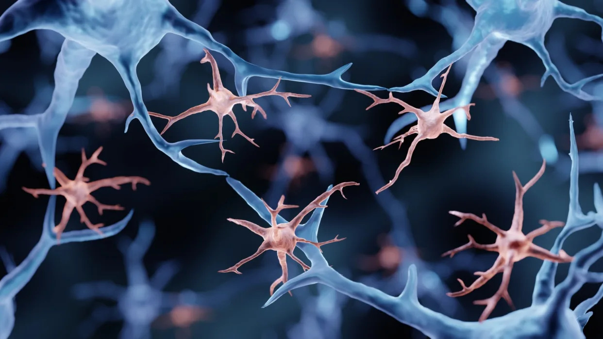
Illustration of microglial cells (in pink) interacting with neurons (in blue) Extreme mood changes associated with Alzheimer’s disease may be partially driven by brain inflammation , new research suggests.
Historically, the prevailing theory for what causes Alzheimer’s disease was that a gradual buildup of abnormal proteins called amyloid-beta and tau in the brain triggers a cascade of events, leading to nerve damage, the death of brain cells, and symptoms of cognitive decline and mood problems.
However, emerging evidence suggests that inflammation in the brain may also be involved in the development of the disease. Specific culprits include immune cells called microglia , which normally promote inflammation in response to injury or disease. Activated microglia have been found to interact with amyloid-beta and tau proteins and may influence the progression of Alzheimer’s disease .
And now, in a study published Nov. 27 in the journal JAMA Network Open , scientists have provided what they say is the first strong evidence that the neuropsychiatric symptoms of Alzheimer’s are directly associated with microglia activation.
Better understanding the role inflammation plays in the development of Alzheimer’s could take us one step closer to developing more targeted treatments for the disease, the researchers say.
Related: Gene variant carried by 1 in 5 people may guard against Alzheimer’s and Parkinson’s, massive study finds
“Neuropsychiatric symptoms such as irritability, agitation, anxiety and depression are among the most difficult symptoms to treat in patients with Alzheimer’s,” Dr. Cristiano Aguzzoli , the study’s lead author and a postdoctoral researcher at the University of Pittsburgh, said in a statement .
“Here, we show for the first time that brain inflammation may be to blame for these symptoms,” he said.
In the new study, the authors recruited 70 people who had no symptoms of cognitive decline and 39 who were cognitively impaired, either displaying early signs of memory loss known as mild cognitive impairment or dementia caused by Alzheimer’s. The participants ranged from age 38 to 87.
The authors assessed whether the participants displayed any of the mood problems that are characteristic of Alzheimer’s. The researchers also scanned the participants’ brains to look for signs of microglial activation, as well as the accumulation of amyloid-beta and tau proteins.
People with cognitive impairment were more likely to have amyloid-beta and tau proteins in their brains. For example, amyloid-beta was found in 79% of those with cognitive impairment compared to 30% without.
However, even after taking these factors into account, the participants with more severe neuropsychiatric symptoms had a greater level of microglial activation and more substantial signs of inflammation than the participants with milder symptoms. This inflammation specifically affected three regions of the outer layer of the brain. Of the mood symptoms, irritability was most strongly associated with microglial activation, followed by nighttime disturbances and agitation.
The authors didn’t directly compare whether participants’ protein buildup was more or less strongly associated with mood problems than their inflammation levels. So they couldn’t determine whether one factor is more influential than the other. However, the researchers think it’s possible that both are playing a role.
In addition to conducting these tests, the researchers asked the caregivers of the patients with cognitive decline to fill out a questionnaire about their experiences. They found that the caregivers were more likely to report distress when looking after participants with greater levels of brain inflammation, particularly when it was linked to symptoms of irritability. Caregivers were also more likely to report that patients experienced rapid mood swings when the patients had high inflammation levels.
Going forward, the authors want to conduct more studies with larger groups of patients, including those who are at later stages of the disease and experience more extreme neuropsychiatric symptoms, such as hallucinations or delusions . This would assess whether the team’s findings are more generalizable to the wider population of people with Alzheimer’s.
RELATED STORIES
— Could vaccines prevent and treat Alzheimer’s disease?
— A man’s rare gene variant may have shielded him from devastating form of early Alzheimer’s
— Brain training probably won’t reduce Alzheimer’s risk — here’s why
In the meantime, they hope this research will act as a springboard for developing new therapies for Alzheimer’s and possibly other types of dementia.
“Since both neuroinflammation and neuropsychological abnormalities are found in several other types of dementia, including Parkinson’s dementia, we are collaborating with scientists around the world to expand these findings to these other diseases,” Dr. Tharick Pascoal , co-senior study author and an associate professor of psychiatry and neurology at the University of Pittsburgh, said in the statement.
This article is for informational purposes only and is not meant to offer medical advice.
Ever wonder why some people build muscle more easily than others or why freckles come out in the sun ? Send us your questions about how the human body works to community@livescience.com with the subject line “Health Desk Q,” and you may see your question answered on the website!
Beyond Buzz: 10 Ways Neurotech Is Helping People In The Real World
Beyond the buzz, neurotechnology is helping people in a growing list of areas spanning clinical, … [+] Fancy headsets to manage ADHD. Bluetooth stickers to predict seizures. Mind controlled games to gauge dementia. And of course, implanted chips to relieve pain, restore movement, and eventually, do a whole lot more.
Welcome to the neurotechnology frontier, where brain activity is the coin of the realm. “Neurotech” is an umbrella category for hardware and software tools—think sensors and wearables, invasive and non, combined with powerful AI modeled on large datasets—that are accelerating our ability to understand and influence brain activity.
Acronyms abound: Various forms of sound, light, magnetics and electricity are helping scientists, clinicians and consumers understand brain health from the inside out (and increasingly, vice versa).
The casual reader would be forgiven for thinking it all felt a bit academic, clinical, technical and complicated, because it is. But neurotech’s moment has arrived: This space is moving fast, with growing interest driven by private and public funding, regulatory reforms and media coverage. So it’s a good time to understand the basics of how neurotech is helping people.
Here are 10 places to start. The Day Before Is Real But Lied About Everything Else
The Game Awards Was A Full On Parody Of Itself This Year
Stroke Recovery
The CDC estimates that someone has a stroke every 40 seconds. Neurotechnology plays a growing role in both diagnosis and treatment. Fast, accurate imaging tools are enabling clinicians to get a clear picture of what’s happened in a person’s brain, enabling prompt treatment. For recovery, neurotech’s ability to send thoughts into hardware is revolutionizing rehab. One company’s FDA-cleared wearable gloves are helping people retrain neuromuscular function for upper limb control. Other startups are creating personalized, wearable-driven platforms to help people tune up their ability to walk by syncing steps to music.
Forbes Daily: Get our best stories, exclusive reporting and essential analysis of the day’s news in your inbox every weekday.
By signing up, you accept and agree to our Terms of Service (including the class action waiver and arbitration provisions), and Privacy Statement . Cognitive Decline
Neurotech is opening avenues into understanding of mild cognitive impairment and Alzheimer’s disease. On the research front, neurotech companies are working on pinpointing biomarkers of cognitive decline by capturing and analyzing multimodal datasets, spanning neurofeedback wearables with camera, microphone, biosensors, and digital cognitive assessments. Other companies are treating symptoms with non-invasive, non-chemical tools, such as targeted light and sound waves delivered through wearable headsets. Multiple Sclerosis
As a disease without a known cure, managing symptoms and understanding progression of MS is a vital aspect of the care journey for patients and caregivers alike. Neurotech startups are syncing up brain activity, motor function, cognitive performance and other inputs with AI for greater precision in tracking symptoms and overall health than periodic paper surveys that yield subjective and variable data. As in stroke rehab, some platforms have published research demonstrating improvements in fine motor control, gait, and speech. Parkinson’s Disease
Parkinson’s was one of the first approved uses of implanted electrodes in a procedure known as deep brain stimulation. A quarter century later, more advanced implants are coming to market, along with new, noninvasive techniques. Imagine you’re laying inside an MRI awaiting a brain scan. Now imagine you’ve got on a helmet inside the chamber containing a precision ultrasound device, controlled remotely by the doctor standing a few feet away using the MRI for real-time visibility, that zaps plaques in your brain. Paralysis
Wearable AR headsets are creating new avenues for communication. Much of the hype about invasive “brain computer interfaces” comes from a certain billionaire’s overfunded company. But the broader class of implanted microprocessor companies represents a nexus of science, technology and medicine. These tend to focus on helping people gain or regain motor function, communication and more. While today’s population for this highly regulated technology remains small, limited to trials of the devices and implanting surgical procedures, “neuroethicists” are actively grappling with a near-future when human-computer symbiosis is available more broadly. Seizure Prediction
The EEG, a staple in diagnosis and monitoring of seizures, turns 100 next year, and disruption has arrived in the multi-billion-dollar epilepsy care market. Portable monitoring kits, disposable bluetooth adhesives, video-synced diagnostics and AI-enabled big data prediction tools are introducing more consumer friendly, remote-enabled care systems. These next-gen sensors are more convenient for patients, and offer a glimpse of innovation in form and function. Traumatic Brain Injuries
From eye-tracking visors that identify signs of concussion to novel wearables incorporating next-generation diagnostics for brain bleeds to clinical decision-support AI powered by tens of thousands of brain scans, there’s never been a better time to bump your head. Kidding. Beyond greater precision in diagnosis, emerging research is also pointing to the benefits of implantable electrodes for people recovering from moderate to severe injuries. Opioid Use Disorder
America’s scourge has generated a proliferation of novel investments to understand and treat addiction, pain, PTSD and other drivers of opioid use disorder. Various neurotech startups funded by public grants from programs like HEAL and SBIR, are building self-management tools, researching brain function and experimenting with treatment of specific symptoms. One example from a stealth mode company is a transcranial ultrasound device using MRI-based brain maps and electroencephalography (EEG) tracking to understand how better sleep can aid the recovery process. Elite Performance
Multimodal sensors (brain function, heart rate, eye tracking, other biometrics) are consolidating into mobile platforms that move with athletes or soldiers, from training labs to the field. Professional athletes and collegiate teams are using such tools to boost hand-eye coordination, cut reaction time and tune physiological aspects of performance under duress. The U.S. military has been one of the earliest and largest funders of applied neurotech research through the Defense Advanced Research Projects Agency (DARPA) and other programs. Myriad applications span security, “lethality,” field diagnostics and interventions, health research, advanced craft piloting and, let’s be real here, plenty of top-secret stuff we’ll never know about. Consumer Productivity
A global landscape of startups […]
Redefining Brain Evolution: Unveiling the “Little Brain’s” Role in the Human Cognitive Leap
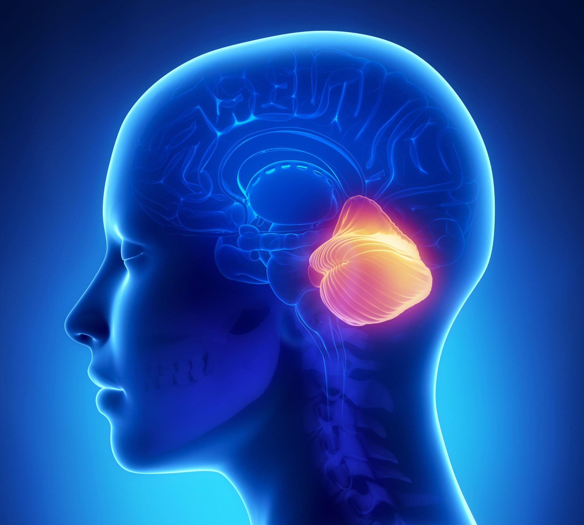
Heidelberg University researchers have mapped the cerebellum’s development in humans, mice, and opossums, uncovering its complex structure and significant role in human cognitive evolution. Their findings offer insights into brain development and diseases, with a focus on Purkinje cells and genetic variations over 160 million years. Scientists at Heidelberg have revealed genetic mechanisms that govern the formation of varied cell types in the human cerebellum and in other mammals.
The advancement of higher cognitive abilities in humans is predominantly associated with the growth of the neocortex, a brain area key to conscious thinking, movement, and sensory perception. Researchers are increasingly realizing, however, that the “little brain” or cerebellum also expanded during evolution and probably contributes to the capacities unique to humans, explains Prof. Henrik Kaessmann from the Center for Molecular Biology of Heidelberg University .
His research team has – together with Prof. Dr Stefan Pfister from the Hopp Children’s Cancer Center Heidelberg – generated comprehensive genetic maps of the development of cells in the cerebella of humans, mice, and opossums. Comparisons of these data reveal both ancestral and species-specific cellular and molecular characteristics of cerebellum development spanning over 160 million years of mammalian evolution. Revealing Cerebellum’s Complexity
“Although the cerebellum, a structure at the back of the skull, contains about 80 percent of all neurons in the whole human brain, this was long considered a brain region with a rather simple cellular architecture,” explains Prof. Kaessmann. In recent times, however, evidence suggesting a pronounced heterogeneity within this structure has been growing, says the molecular biologist. Genetic maps of the development of cells in the cerebella of humans, mice, and opossums shed light on ancestral and species-specific cellular and molecular characteristics of cerebellum development. Credit: Mari Sepp The Heidelberg researchers have now systematically classified all cell types in the developing cerebellum of humans, mice, and opossums. To do so they first collected molecular profiles from almost 400,000 individual cells using single-cell sequencing technologies. They also employed procedures enabling spatial mapping of the cell types. Purkinje Cells and Cognitive Function
On the basis of these data the scientists noted that in the human cerebellum, the proportion of Purkinje cells – large, complex neurons with key functions in the cerebellum – is almost double that of mouse and opossum in the early stages of fetal development. This increase is primarily driven by specific subtypes of Purkinje cells that are generated first during development and likely communicate with neocortical areas involved in cognitive functions in the mature brain.
“It stands to reason that the expansion of these specific types of Purkinje cells during human evolution supports higher cognitive functions in humans,” explains Dr Mari Sepp, a postdoctoral researcher in Prof. Kaessmann’s research group “Functional evolution of mammalian genomes.” Genetic Analysis and Evolutionary Insights
Using bioinformatic approaches, the researchers also compared the gene expression programs in cerebellum cells of humans, mice, and opossums. These programs are defined by the fine-tuned activities of a myriad of genes that determine the types into which cells differentiate in the course of development. Genes with cell-type-specific activity profiles were identified that have been conserved across species for at least about 160 million years of evolution.
According to Henrik Kaessmann, this suggests that they are important for fundamental mechanisms that determine cell type identities in the mammalian cerebellum. At the same time, the scientists identified over 1,000 genes with activity profiles differing between humans, mice, and opossums.
“At the level of cell types, it happens fairly frequently that genes obtain new activity profiles. This means that ancestral genes, present in all mammals, become active in new cell types during evolution, potentially changing the properties of these cells,” says Dr Kevin Leiss, who – at the time of the studies – was a doctoral student in Prof. Kaessmann’s research group. Implications for Biomedical Research
Among the genes showing activity profiles that differ between humans and mice – the most frequently used model organism in biomedical research – several are associated with neurodevelopmental disorders or childhood brain tumors, Prof. Pfister explains. He is a director at the Hopp Children’s Cancer Center Heidelberg, heads a research division at the German Cancer Research Center, and is a consultant pediatric oncologist at Heidelberg University Hospital.
The results of the study could, as Prof. Pfister suggests, provide valuable guidance in the search for suitable model systems – beyond the mouse model – to further explore such diseases.
Reference: “Cellular development and evolution of the mammalian cerebellum” by Mari Sepp, Kevin Leiss, Florent Murat, Konstantin Okonechnikov, Piyush Joshi, Evgeny Leushkin, Lisa Spänig, Noe Mbengue, Céline Schneider, Julia Schmidt, Nils Trost, Maria Schauer, Philipp Khaitovich, Steven Lisgo, Miklós Palkovits, Peter Giere, Lena M. Kutscher, Simon Anders, Margarida Cardoso-Moreira, Ioannis Sarropoulos, Stefan M. Pfister and Henrik Kaessmann, 29 November 2023, Nature .
DOI: 10.1038/s41586-023-06884-x
Also participating in the studies – apart from the Heidelberg scientists – were researchers from Berlin as well as China, France, Hungary, and the United Kingdom. The European Research Council financed the research. The data are available in a public database.