Learn about brain health and nootropics to boost brain function
Researchers discover that the spinal cord works with the brain to control complex motor functions


(Natural News) The spinal cord displays more capability than most people give it credit for. A new study reported that this structure directly controls complicated motor functions in the arm.
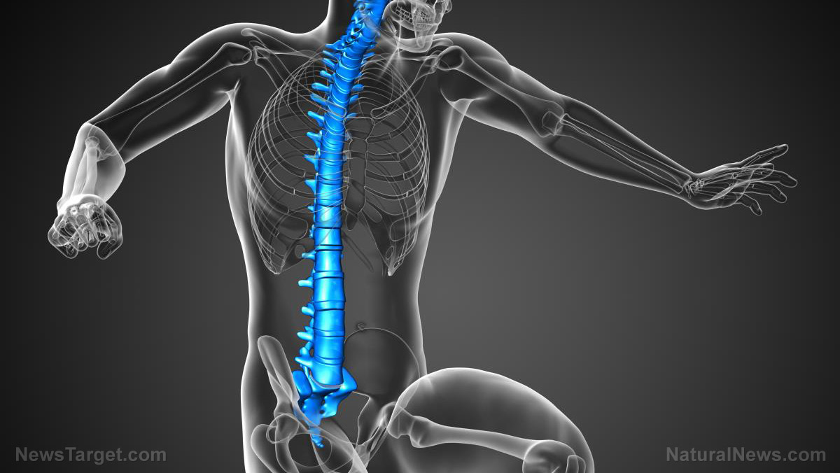
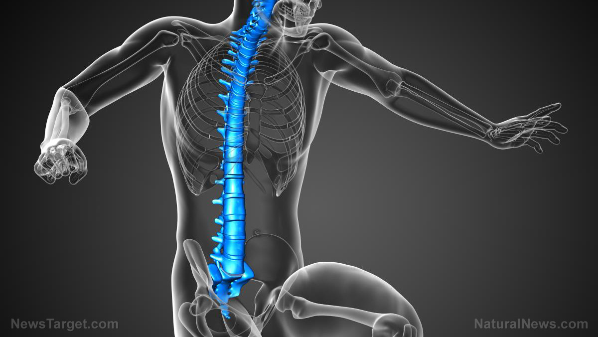
The spinal cord runs down the length of the spine. It is a large and important part of the central nervous system. In humans, its neural circuits govern the pain reflex. In animals, the spinal cord controls simple motor functions.
Western University researchers reported that the spinal cord handles higher motor functions as well. For example, it controls the positioning of the hand in a space – a simple task that is more complicated than it sounds.
“This research has shown that a least one important function is being done at the level of the spinal cord and it opens up a whole new area of investigation to say, ‘what else is done at the spinal level and what else have we potentially missed in this domain?'” asked Western researcher Dr. Andrew Pruszynski.
As senior researcher and supervisor of the Western team, Pruszynski oversaw the investigation of the human spinal cord. The findings of their study were published in the journal Nature Neuroscience. (Related: Curcumin performs BETTER than drugs and surgery at treating spinal cord injuries – review.)
Robotic exoskeleton helps researchers determine what controls the hand
Manipulation of the positioning of a hand relies on sensory data from several nearby joints. The elbow and wrist, in particular, provide most of the data needed to determine the location of the appendage.
100% organic essential oil sets now available for your home and personal care, including Rosemary, Oregano, Eucalyptus, Tea Tree, Clary Sage and more, all 100% organic and laboratory tested for safety. A multitude of uses, from stress reduction to topical first aid. See the complete listing here, and help support this news site.
Previously, researchers believed that sensory data is processed by the cerebral cortex of the brain. They thought the brain also sends motor commands to the hand.
The Western University research team availed of special robotic systems at the university’s Brain and Mind Institute. They used a robotic exoskeleton with three degrees of freedom of movement, which were worn by volunteers.
In the experiment, the participants were instructed to hold their hand in a specified position. Next, the exoskeleton bumped the hand away from the location with a concurrent movement of the elbow and wrist.
The researchers recorded the period it took for the muscles in the elbow and wrist of the volunteers to react to the robot knocking their hand away. They also noted whether or not the reactions succeeded in returning the disturbed hand back to the required location.
Finally, they evaluated the latency of the reaction. A short lapse indicated a faster trip to the spinal cord, while a longer lag time meant that the data went all the way to the brain.
The spinal cord can perform more complex motor functions than previously believed
“We found that these responses happen so quickly that the only place that they could be generated from is the spinal circuits themselves,” explained Western University researcher Dr. Jeff Weiler. “What we see is that these spinal circuits don’t really care about what’s happening at the individual joints, they care about where the hand is in the external world and generate a response that tries to put the hand back to where it came from.”
The spinal cord swiftly reacted to certain sensory inputs through what was called a “stretch reflex.” The researchers believe that these spinal reflexes returned the triggered muscle to their previous length before the stretch took place.
The findings of the Western University experiment indicated that the stretch reflexes did more than just restore muscles to their earlier state. The researchers concluded that the spinal cord directly controlled the hand and made sure it stayed in the right place. They believed that their discovery might lead to new physical rehabilitation therapies that involve the spinal cord.
Sources include:
Click here to view full article