Nature Knows and Psionic Success
God provides
This Is The Largest Map of The Human Brain Ever Made
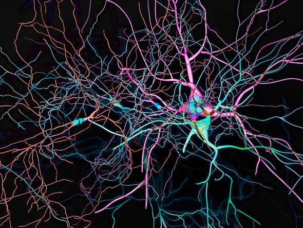
Insights into thousands of types of brain cell could improve understanding of diseases and cognition. Credit: Researchers have created the largest atlas of human brain cells so far, revealing more than 3,000 cell types — many of which are new to science. The work, published in a package of 21 papers today in Science , Science Advances and Science Translational Medicine , will aid the study of diseases, cognition and what makes us human, among other things, say the authors.
The enormous cell atlas offers a detailed snapshot of the most complex known organ. “It’s highly significant,” says Anthony Hannan, a neuroscientist at the Florey Institute of Neuroscience and Mental Health in Melbourne, Australia. Researchers have previously mapped the human brain using techniques such as magnetic resonance imaging, but this is the first atlas of the whole human brain at the single-cell level, showing its intricate molecular interactions, adds Hannan. “These types of atlases really are laying the groundwork for a much better understanding of the human brain.”
The research is part of the US National Institutes of Health’s Brain Research through Advancing Innovative Neurotechnologies Initiative — Cell Census Network (BICCN), a collaboration between hundreds of scientists. The programme’s goals include cataloguing brain cell types across humans, non-human primates and mice to improve understanding of the cellular mechanisms behind poorly understood brain disorders. The data from the 21 studies have been made publicly available on the Neuroscience Multi-omic Archive online repository. Cellular menagerie
Kimberly Siletti, a neuroscientist now at the University Medical Center Utrecht in the Netherlands, and her team laid the cornerstone for the atlas by sequencing the RNA of more than 3 million individual cells from 106 locations covering the entire human brain, using tissue samples from three deceased male donors. They also included one motor cortex dissection from a female donor that had been used in previous studies. Their analysis documented 461 broad categories of brain cell that included more than 3,000 subtypes. “I was surprised at how many different cell types there were,” says Siletti.
Neurons — cells in the brain and nervous system that send and receive signals — varied widely in different parts of the brain, suggesting different functions and developmental histories. The mix of neurons and other cell types also differed across each region; some cells were only found in specific locations. The brainstem — a relatively under-studied structure connecting the brain to the spinal cord — harboured a particularly high number of neuron types, says study co-author Sten Linnarsson, a molecular systems biologist at the Karolinska Institute in Stockholm, Sweden. “One of the big surprises here is how incredibly complex the brainstem is.”
Other studies drilled into the mechanisms of gene regulation and expression in different cells. Joseph Ecker, a molecular biologist at the Salk Institute for Biological Studies in La Jolla, California, and his colleagues investigated the brain through an epigenetic lens using tissue samples from the same three donors. They analysed chemical markers that switch genes on or off in more than 500,000 individual cells. The various molecules that acted as switches enabled the team to identify nearly 200 brain cell types. Even the same gene in the same type of cell could have different characteristics across the brain. One gene was turned on with one switch at the front of the brain and with another at the back. “There are remarkable regional differences,” says study co-author Wei Tian, a computational biologist at the Salk Institute.
Pinpointing the switches that activate or block gene expression in brain cells could be useful for diagnosing brain disorders and developing tailored treatments, says Ecker. “That’s another tool that comes out of the toolbox we’re building,” he says. Disease risk
Improving understanding of how genetic switches might contribute to disease risk was also a focus for Bing Ren, a molecular biologist at the University of California, San Diego, and his team. They analysed how more than one million brain cells from the three donors access and use genetic information. The researchers uncovered links between certain brain cell types and neuropsychiatric disorders, including bipolar disorder, depression and schizophrenia.
Ren and his colleagues used the cell-type data to predict how the genetic switches influence gene regulation and increase the risk of neurological diseases. For instance, in cells called microglia , which clear away dead or damaged cells, the presence of some genetic switches was strongly linked to risks of Alzheimer’s disease. Such findings can be used to test whether particular genes or faulty switches contribute directly to the onset of disease. “This is made possible because we have — for the first time — delineated the genetic switches for hundreds of different cell types,” says Ren.
The next step for the BICCN team is to sequence more cells from all parts of the brain, says Ren. The researchers will also work with more tissue samples to build a picture of how the human brain can vary across populations and age groups. “This is only the beginning,” says Ren.
This article is reproduced with permission and was first published on October 12, 2023. Sign up for Scientific American ’s free newsletters. ABOUT THE AUTHOR(S)
Read more at www.scientificamerican.com

The Power Within by Corey Daniels book available for only $2.99
Mind has both conscious and subconscious halves. These are likened to a driver and the truck he drives. The driver plans the destination and observes road conditions, while the truck provides motive power. Your subconscious mind is like the truck and it only goes in the direction in which is a steered. This can be the road or off a cliff. Likewise, the consciousness paints a picture of what the world is and what your goals are and the subconscious acts on them through emotion, physical response and energy, whether these are correct, rational images or false negative ones. The subconscious is also like an emotional reservoir which your body and mind draw responses from to external stimuli.
The Power Within lays out a method of programming your subconscious and tapping into the Holy Spirit, God voice or what the Greeks called the daimon (godman).