Nature Knows and Psionic Success
God provides
How Brain Flexibility Emerges in Infants
Summary: Neuroimaging study reveals how human brain complexity and cognitive flexibility develops during early infancy.
Source: University of North Carolina Health Care
Cognitive flexibility refers to the ability to readily switch between mental processes in response to external stimuli and different task demands. For example, when our brains are processing one task, an external stimulus is present, requiring us to switch our mental processes to attend to this external stimulus. This ability of switching from one to another mental task is the cognitive flexibility. Such flexibility can predict reading ability, academic success, resilience to stress, creativity, and lower risk of various neurological and psychiatric disorders. To shed light on the development of this critical cognitive process during early infancy, researchers at the UNC Biomedical Research Imaging Center (BRIC) at the UNC School of Medicine conducted a brain imaging study in infants to examine the emergence of neural flexibility, which refers to the frequency with which a brain region changes its role (or allegiance to one functional network to another). Neural flexibility is thought to underlie cognitive flexibility.
Publishing their work in the Proceedings of the National Academy of Sciences (PNAS), the researchers show that brain regions with high neural flexibility appear consistent with the core brain regions that support cognitive flexibility processing in adults, whereas brain regions governing basic brain functions, such as motor skills, exhibit lower neural flexibility in adults, demonstrating the emergence of functionally flexible brains during early infancy.
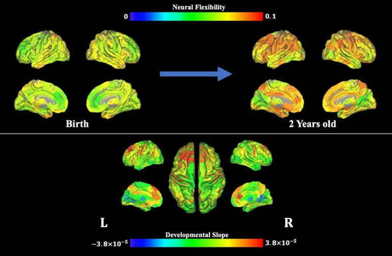
For this study, the authors used magnetic resonance imaging to examine brain activity up to seven times in 52 typically developing infants under the age of two during natural sleep. The researchers, led by Weili Lin, PhD, director of BRIC, the Dixie Lee Boney Soo Distinguished Professor of Neurological Medicine, and Vice Chair of Basic Research in the UNC Department of Radiology, found that neural flexibility increased with age across the whole brain, and specifically in brain regions that control movement, potentially enabling infants to learn new motor skills. Neural flexibility also increased with age in brain regions involved in higher-level cognitive processes, such as attention, memory, and response inhibition, indicating continuing development of these functional networks as babies become toddlers.
The age-related increase in neural flexibility was highest in brain regions already implicated in cognitive flexibility in adults, suggesting that cognitive flexibility may start to develop during the first two years of life.
“Neural flexibility in these brain regions may reflect early developmental processes that support the later emergence of cognitive flexibility,” Lin said. “What we’ve imaged, in essence, is the brain’s flexibility setting the stage for later maturity of higher cognitive brain functions.”
Additional analysis of brain regions with especially high neural flexibility revealed the presence of relatively weak and unstable connections from these regions to other parts of the brain, potentially showing how these regions can rapidly switch their allegiances between different functional networks. By contrast, neural flexibility in brain regions involved in visual functions remained relatively low throughout the first two years of life, suggesting that these regions had already matured. This image is of MRI data, showing neural flexibility over time. Image is credited to Biomedical Research Imaging Center at UNC-Chapel Hill. Lower levels of neural flexibility (i.e., greater established brain maturity) of visual brain regions at three and 18 months of age were associated with better performance on cognitive and behavioral assessments at the age of five or six years.
These findings provide insights into the development of higher-level brain functions, and could be used to predict cognitive outcomes later in life. The developed approach of assessing neural flexibility non-invasively could also provide a new means to assess subjects with neurodevelopmental disorders.
First author of this study is BRIC postdoctoral researcher Weiyan Yin, PhD. Other authors aside from Yin and Lin include Sheng-Che Hung, MD, clinical instructor of radiology; Han Zhang, PhD, assistant professor of radiology; Li Wang, PhD, research assistant professor of radiology; Dinggang Shen, PhD, Director of the UNC BRIC Image Analysis Core and professor of radiology, Hongtu Zhu, PhD, Peter Mucha, PhD, professor of mathematics and applied physical sciences; and Jessica Cohen, PhD, assistant professor of psychology and neuroscience, all at UNC-Chapel Hill. All but Mucha are members of BRIC.
Funding: The National Institutes of Health funded this work.
About this neurodevelopment research article
Source:
University of North Carolina Health Care
Contacts:
Mark Derewicz – University of North Carolina Health Care
Image Source:
The image is credited to Biomedical Research Imaging Center at UNC-Chapel Hill.
Original Research: Open access
“The emergence of a functionally flexible brain during early infancy” by Weili Lin et al. PNAS .
Abstract
The emergence of a functionally flexible brain during early infancy
Adult brains are functionally flexible, a unique characteristic that is thought to contribute to cognitive flexibility. While tools to assess cognitive flexibility during early infancy are lacking, we aimed to assess the spatiotemporal developmental features of “neural flexibility” during the first 2 y of life. Fifty-two typically developing children 0 to 2 y old were longitudinally imaged up to seven times during natural sleep using resting-state functional MRI. Using a sliding window approach, MR-derived neural flexibility, a quantitative measure of the frequency at which brain regions change their allegiance from one functional module to another during a given time period, was used to evaluate the temporal emergence of neural flexibility during early infancy. Results showed that neural flexibility of whole brain, motor, and high-order brain functional networks/regions increased significantly with age, while visual regions exhibited a temporally stable pattern, suggesting spatially and temporally nonuniform developmental features of neural flexibility. Additionally, the neural flexibility of the primary visual network at 3 mo of age was significantly and negatively associated with cognitive ability evaluated at 5/6 y of age. The “flexible club,” comprising brain regions with neural flexibility significantly higher than whole-brain neural flexibility, were consistent with brain regions known to govern cognitive flexibility in adults and exhibited unique characteristics when compared to the functional hub and diverse club regions. Thus, MR-derived […]
Read more at neurosciencenews.com

The Power Within by Corey Daniels book available for only $2.99
Mind has both conscious and subconscious halves. These are likened to a driver and the truck he drives. The driver plans the destination and observes road conditions, while the truck provides motive power. Your subconscious mind is like the truck and it only goes in the direction in which is a steered. This can be the road or off a cliff. Likewise, the consciousness paints a picture of what the world is and what your goals are and the subconscious acts on them through emotion, physical response and energy, whether these are correct, rational images or false negative ones. The subconscious is also like an emotional reservoir which your body and mind draw responses from to external stimuli.
The Power Within lays out a method of programming your subconscious and tapping into the Holy Spirit, God voice or what the Greeks called the daimon (godman).