Nature Knows and Psionic Success
God provides
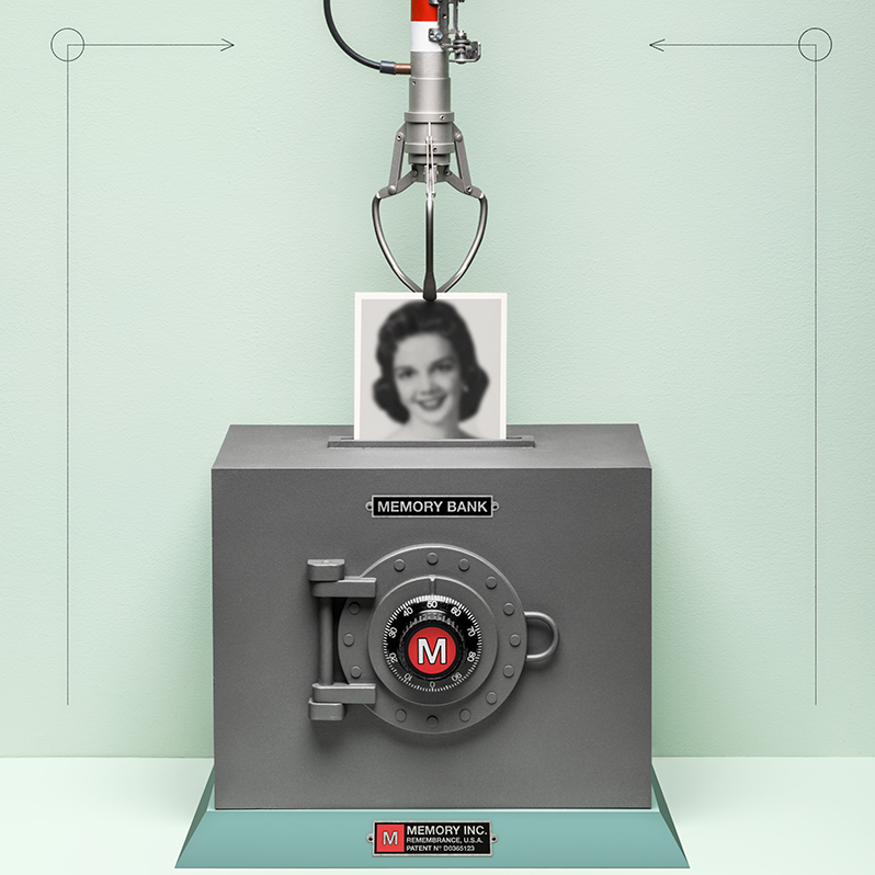
Living Memory
It is possible to watch a memory forming in the brain. Researcher Sheena Josselyn at the Hospital for Sick Children in Toronto, for instance, looks through an ultralight “mini endoscope” attached to a lens in the brain of a mouse. The animal’s brain cells have been tagged with substances that make them light up when they become active, and she waits to see exactly which neurons “switch on” in response to an event she thinks the animal will remember—most often, a mild electric shock. Josselyn is fascinated by fundamental questions about memory, including how individual neurons “decide” to become part of a particular memory, called an engram. In a landmark experiment, published in 2009 in Science, she noted the role of neurons’ “excitability.” At any given time, certain neurons show an increased readiness to pass along electrical signals and connect with other neurons. Using a genetic tweak in mice to increase levels of a protein called CREB, she was able to increase the excitability of particular neurons, and found that thisinfluenced where in the brain a memory was encoded. She showed that the CREB-boosted cells were nearly four times as likely as neighboring neurons to become part of the engrams that her team prompted. The memory in that experiment was an association between a shock and a musical tone. To prove that the neurons actually encoded this, she turned off the brain cells that had lit up during memory formation, selectively killing them with a toxin. When exposed to the sound again, the mouse appeared to forget what it had learned, no longer freezing in anticipation of a shock. Josselyn has conducted a host of similar experiments, both refining her team’s ability to map engrams and devising new techniques to switch them off and on again. Learning how to […]
Click here to view full article

The Power Within by Corey Daniels book available for only $2.99
Mind has both conscious and subconscious halves. These are likened to a driver and the truck he drives. The driver plans the destination and observes road conditions, while the truck provides motive power. Your subconscious mind is like the truck and it only goes in the direction in which is a steered. This can be the road or off a cliff. Likewise, the consciousness paints a picture of what the world is and what your goals are and the subconscious acts on them through emotion, physical response and energy, whether these are correct, rational images or false negative ones. The subconscious is also like an emotional reservoir which your body and mind draw responses from to external stimuli.
The Power Within lays out a method of programming your subconscious and tapping into the Holy Spirit, God voice or what the Greeks called the daimon (godman).